

Scientifica's top 10 articles of 2018
In 2018, a number of exciting neuroscience research articles were published. We also released a variety of articles and videos to help support you with your research. Here’s Scientifica’s collection of the top 10 articles from 2018.
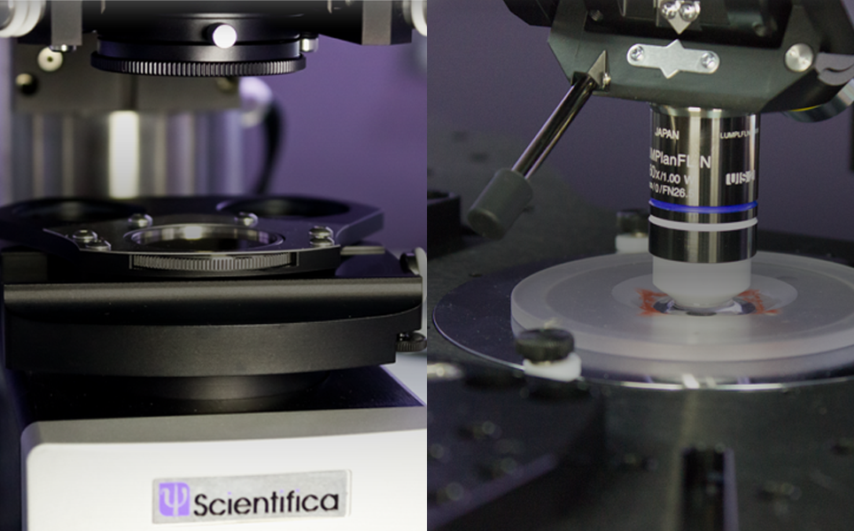
1. #LabHacks: Tips for cleaning the optics of your microscope
Keeping the optical system of your microscope clean at all times is essential for high quality imaging. It is really frustrating if dirt or dust particles are visible when imaging your specimen. If dust spots are left on optical glasses such as lenses, condensers and filters, they can become hard and may attract moisture, further compounding the problem.
To ensure your microscope stands the test of time, cleaning the lenses should be made a regular part of the maintenance routine. Here are some hints and tips that we compiled alongside Olympus UK’s Life Science team, which explain the procedure for cleaning the optical lenses of your microscope.
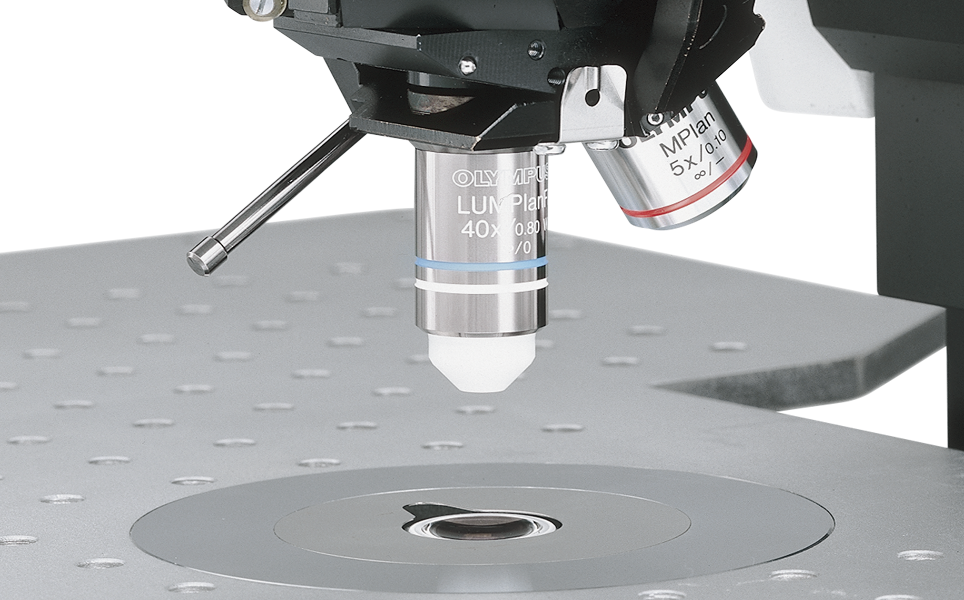
Keep your optics clean
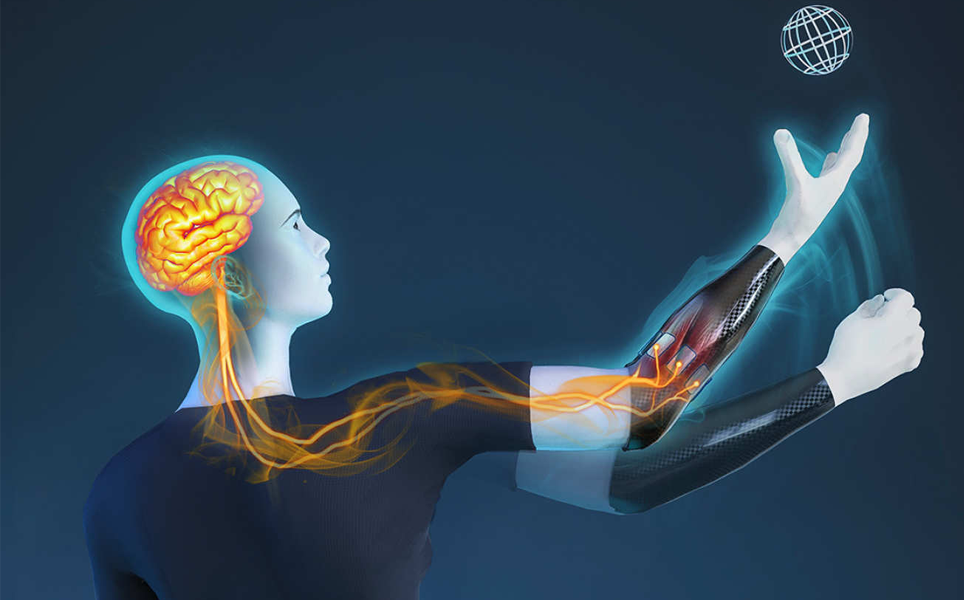
2. Self-learning bionic hand could spark ‘new generation’ of prosthetic limbs
Scientists at Imperial College London and the University of Gottingen have developed a bionic hand that interprets patients’ intentions and moves in the way they want it to.
The bionic limb contains eight electrodes that detect weak electrical signals from the patient’s stump. The signals are then amplified before being sent to a mini computer within the prosthetic limb. The mini-computer interprets the signals and commands the hand to move. Patients found that the limb moved more naturally than other bionic limbs.
Self-learning limb
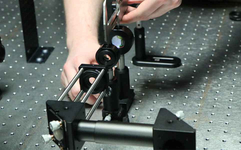
3. #LabHacks: How to align your laser for two-photon imaging
Two-photon microscopy uses a laser to excite fluorescent molecules (fluorophores) within a sample through emitting short pulses of light at high power. Benefits of using two-photon excitation include specifically exciting fluorophores only in the actual focal volume and reduced phototoxicity and photobleaching. Two photon microscopy enables excitation of fluorophores deeper in tissue, as the light used is a longer wavelength so can penetrate deeper into tissue.
This step-by-step guide demonstrates how to safely align a laser for two photon microscopy.
Learn how
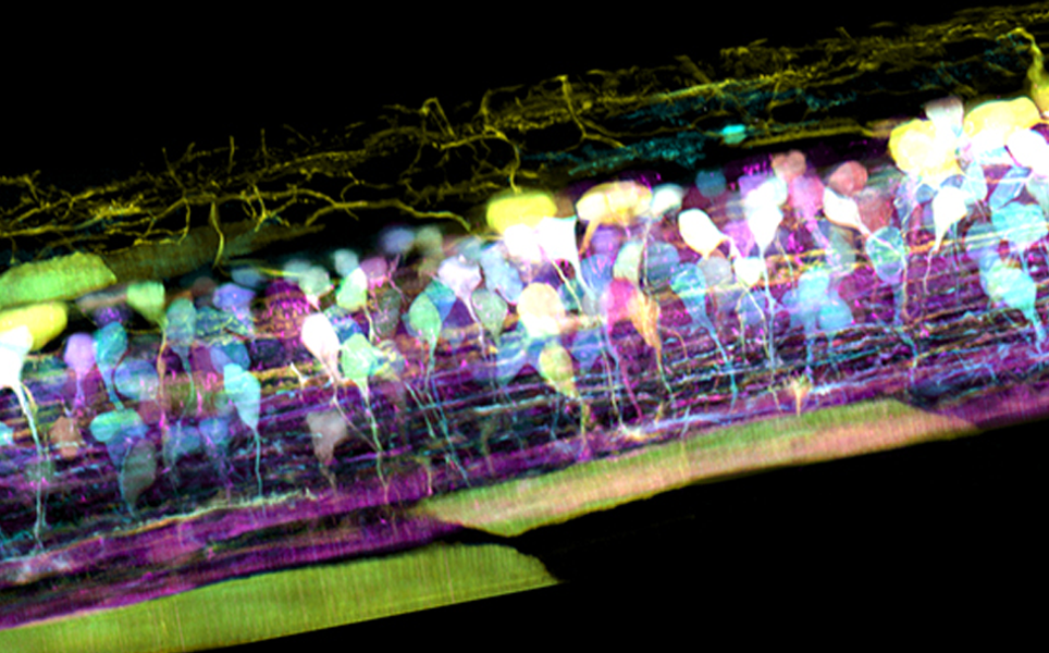
4. New microscope captures detailed 3D movies of cells deep within living systems
Scientists at the Howard Hughes Medical Institute have combined lattice light sheet microscopy with adaptive optics to capture 3D movies of cells within living systems
The new system is fast enough to visualise 3D cellular processes in detail and in real time. It is also non-invasive so does not cause photobleaching and cellular damage, unlike traditional microscopes. The microscope has been able to visualise the following processes in-vivo: clathrin-mediated endocytosis; organelle dynamics in the zebrafish brain and spinal cord neural circuit development, to name a few.
3D movies here
5. Three-photon imaging: How it works
Three-photon imaging is a fluorescence microscopy technique that enables deeper imaging compared to two-photon or one-photon fluorescence microscopy.
Similar to two-photon imaging, where two photons simultaneously interact with a fluorescent molecule, in three-photon imaging, three-photons simultaneously interact with a fluorescent molecule to excite it to a higher electronic state.
Find out more about the advantages and limitations of three-photon imaging and how this imaging method works.
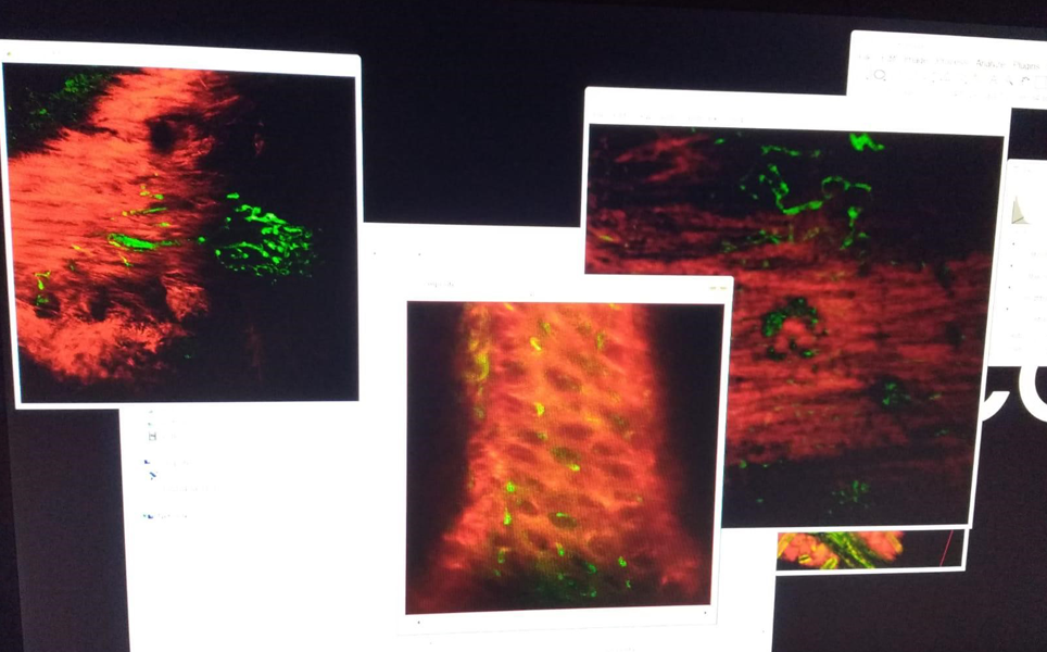
Learn about 3P!
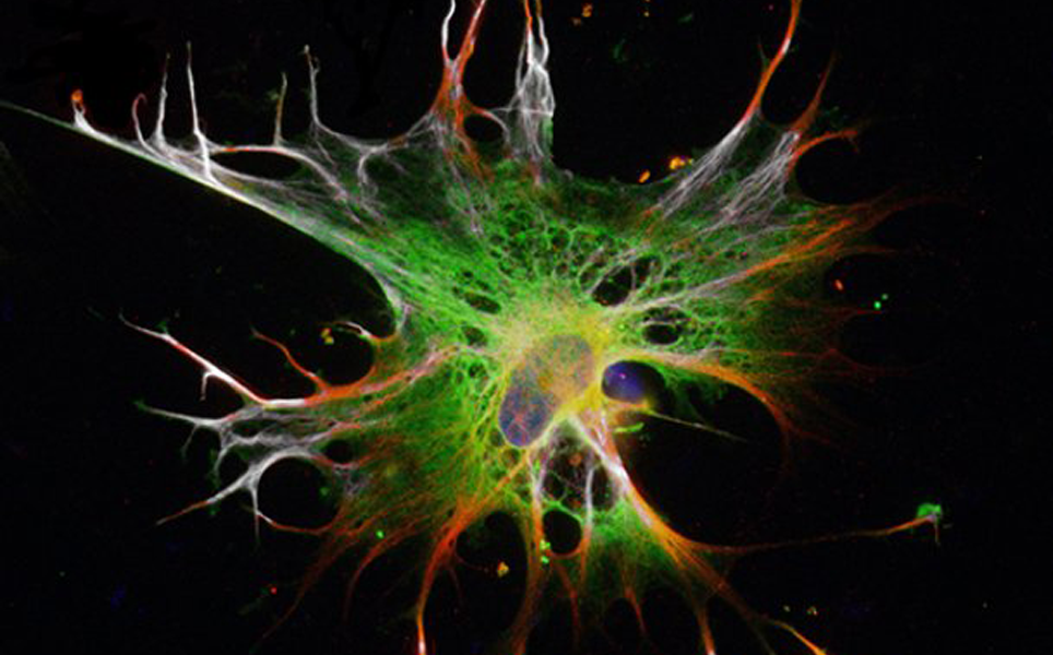
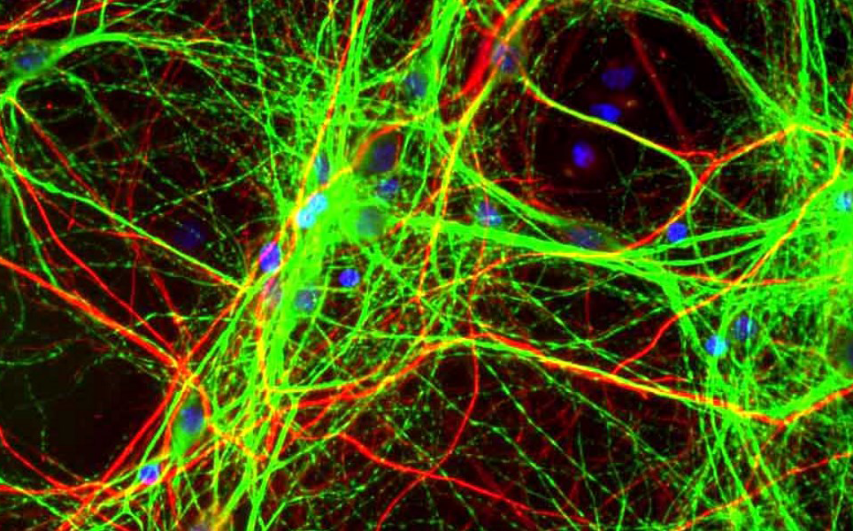
6. New method grows brain cells in two weeks
Researchers at Lund University have found a way to develop astrocytes from embryonic stem cells in just two weeks, compared to it previously taking two months.
Astrocytes have important roles in a variety of brain disorders, including dementia and ALS. The method means that large amounts of functional astrocytes can now be produced in a short amount of time, making it easier to study them.
Find out more
7. A guide to Differential Interference Contrast
Differential Interference Contrast (DIC) is a microscopy technique that introduces contrast to images of specimens which have little or no contrast when viewed using brightfield microscopy. The images produced using DIC have a pseudo 3D-effect, making the technique ideal for electrophysiology experiments.
This article explains the applications of DIC, how it works and how to set it up. A link to a downloadable PDF guide is also included to print off and use in the lab.
More about DIC
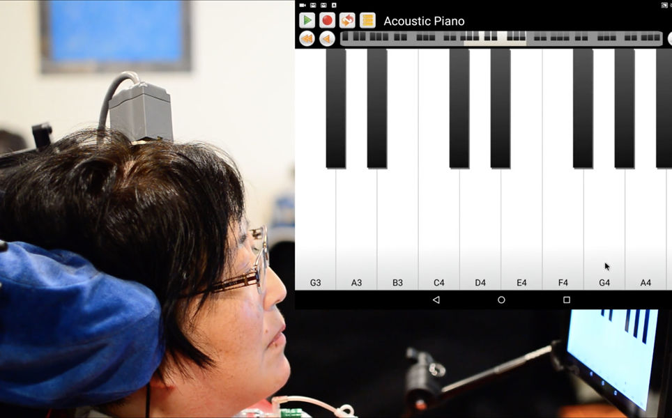
8. Brain-computer interface enables people with paralysis to control tablet devices
In a BrainGate clinical trial, which includes Brown University researchers, a brain-computer interface was found to enable people with paralysis to operate a tablet device by just thinking about making cursor movements and clicks.
The brain-computer interface records neural activity through a small sensor that is placed in the motor cortex. The sensor detects signals associated with intended movements, decodes the signals and sends them to an external device, which in this case was a virtual mouse. This enabled the three clinical trial participants, who had tetraplegia, use a variety of commonly used tablet functions, including email, chat and music-streaming.
Read more
9. Choosing the best light source for your fluorescence experiment
There are a variety of light sources that can be used to illuminate a sample during widefield fluorescence microscopy experiments. Choosing the most suitable one requires evaluating the properties of the light sources and balancing out their pros and cons.
Here, we introduce the three commonly used light sources and discuss their properties as well as their advantages and limitations, to help you make an informed decision of which source is best-suited to your experiment.
The best light source for you
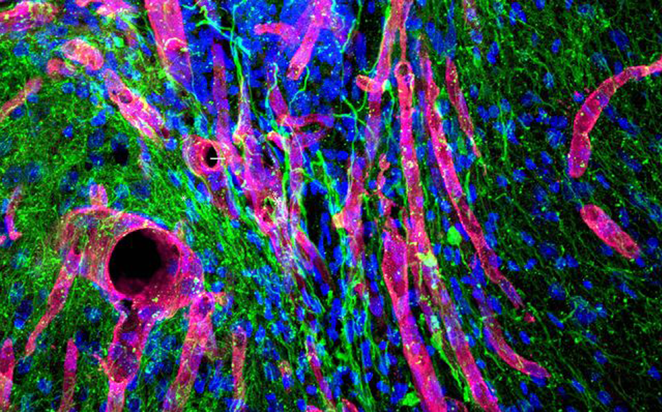
10. Biomaterial developed at UCLA helps regrow brain tissue after stroke in mice
Scientists at UCLA have created a hydrogel that successfully helped neurons and blood vessels regrow in the brains of mice that had been damaged by strokes. Once injected into the site of injury, the gel thickens to create a scaffold for blood vessels and neurons to grow.
The results suggest that this approach could one day be used to treat people who have suffered from a stroke.
Learn more
We hope you enjoyed these as much as we did. We are looking forward to more exciting research in 2019.
Sign up to receive our latest news
Find out about Scientifica's latest product releases, company news, and developments through a range of news articles, customer interviews and product demonstration videos.
)