

Three-photon imaging: How it works
Three-photon imaging is a fluorescence microscopy technique that enables deeper imaging compared to two-photon or one-photon fluorescence microscopy.
Similar to two-photon imaging, where two photons simultaneously interact with a fluorescent molecule, in three-photon imaging, three-photons simultaneously interact with a fluorescent molecule to excite it to a higher electronic state.
The three photons that excite a fluorophore will have longer wavelengths and lower energy than those used to excite the same fluorophore in one- or two-photon fluorescence imaging, for example excitation of green fluorescent protein (GFP) requires:
- 1 photon with a wavelength of 480nm
- 2 photons each with a wavelength of 910nm
- 3 photons each with a wavelength of 1300nm
Advantages
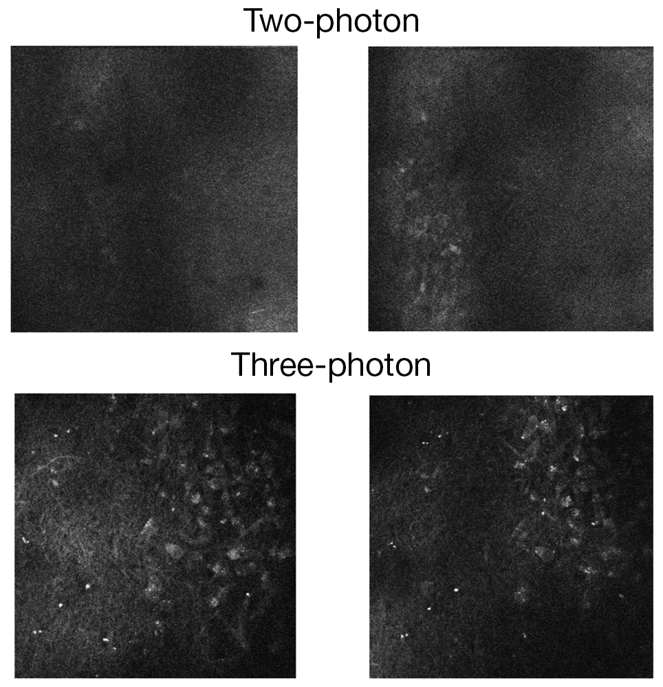
Three-photon imaging is ideal for imaging deeper in scattering tissue, or when imaging through thin scattering layers. Due to the light having a longer wavelength, it is able to penetrate deeper into tissue. The light scatters less, enabling clearer images of structures deep in scattering tissue to be obtained. Fluorophores deeper in tissue can be excited, and, as with other optical sectioning techniques, structures can be visualized in 3D.
How it works
To achieve three-photon excitation, the three photons need to interact with the fluorophore simultaneously (i.e. within less than 1 femtosecond of each other). Therefore, light with a high photon density needs to be used. This is achieved by using fast laser pulses that have high peak power.
A special high-powered laser source is required to achieve the higher photon density and longer wavelengths required for excitation by three photons. The commonly used approach is based on a high power fibre laser pumping an optical parametric amplifier (OPA), which takes light of one wavelength and turns it into light of two different wavelengths. One of these beams has a shorter wavelength than the initial pumped laser. This is the signal beam and can be used for two-photon imaging. The second beam has a longer wavelength than the initial pumped laser. This is the idler beam and is ideal for three-photon imaging.
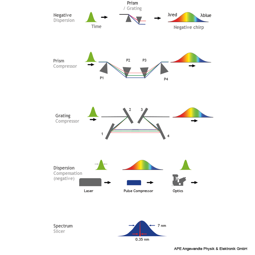
The idler beam is passed through a pre-chirper (pulse compressor), to ensure the shortest possible pulses at the sample.
The pre-chirper achieves the shortest possible pulses at the sample by introducing a negative chirp into the idler beam. Laser pulses are stretched by dispersion of optical material when they pass through optical components such as lenses. This is because the different wavelengths that make up the laser light travel at different speeds through the optical material. Introducing a negative chirp counteracts this effect, so that when the laser pulse goes through glass, the different wavelengths leave the glass at the same time. This is known as dispersion compensation and creates a short pulse with a high photon density.
Read more about pulse compression and dispersion compensation on APE’s website
Limitations
Due to a higher density of photons, and therefore more light being used in three-photon imaging, photodamage and photobleaching are more likely to occur compared to two-photon imaging. Care needs to be taken to keep excitation powers at a biocompatible level.
The main limitation of three-photon imaging is that there is no ability for resonant imaging, due to the very low repetition rate of the laser.
More information about three-photon imaging
If you would like further advice or guidance, please do not hesitate to contact the team at Scientifica.
Find out about Scientifica's latest product releases, company news, and developments through a range of news articles, customer interviews and product demonstration videos.