

Tips for picking a good cell
by Nour Al-muhtasib, PhD
Introduction
Slice electrophysiology is a fickle technique. The success of a day’s experiment hinges on a variety of factors ranging from the cutting solution used to the grounding of the rig. One key to success lies in knowing how to choose the right cells for recording and is an important skill to develop. While some part of the art of picking a healthy cell can be learned, a good part of it just comes with experience.
It all begins with the slice
Finding a good cell starts with having optimal slice quality. The quality of slices depends on factors such as the tissue type, brain region, age of the animal, slice angle (so that dendrites remain intact) and specific cells targeted. While recording from younger animals and certain brain regions may be easier, there are techniques to optimize slices even in challenging conditions. Tips on how to record from adult animals can be found here.
Some general tips include making sure your cutting solution is the optimal temperature, that your vibratome is functioning properly with minimal Z-axis vibration, and that you are using the correct cutting solution for your experiment.
If you mess up slicing or solutions, it would be best to start over. It is a better use of your time to start over than looking through your slices for a single “healthy” cell. Chances are even if that single cell looks healthy, it probably won’t give good reliable data.
Optics. Optics. Optics
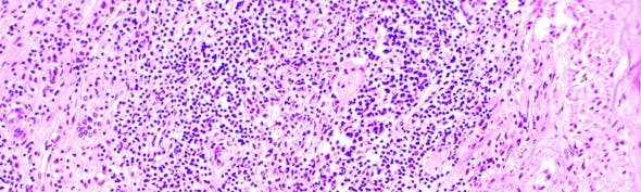
Proper optics are crucial for a successful experiment. Enhanced optics improve the ability to discern health cells. Avoid cells with sharp edges, bloated cells, ones that are super-highly contrasted, or are not contrasted enough. Keep in mind that cells will look different depending on a variety of factors including brain region and age of the animal. For example, pyramidal cells should look pyramidal in shape, not round. If you are using fluorescence, cells that are too bright or have too much somatic fluorescence are likely unhealthy.
Optics will also vary between rigs and the ideal optical setup will depend on your cell/tissue type. While IR-DIC with a 900 nm is ideal for most tissue types, older and heavily myelinated tissue may require alternative methods such as oblique imaging or Dodt gradient contrast. These choices of imaging modality will affect cell identification (For more, see here). The more familiar you become with a rig, the better you become at choosing healthy cells.

Selecting the right electrode size
Choosing the correct electrode size is crucial. The resistance of the electrode is inversely proportional to the tip size. In general, the smaller the cell, the smaller the electrode tip and the higher the pipette resistance. Recording from granule cells with electrodes <5 Mohms can be difficult while large cortical pyramidal neurons can be recorded with 2-3 Mohm electrodes. It is generally easier to achieve seals with higher resistance electrodes. The choice of electrode is dependent upon your recording goals. Voltage clamp recording of large currents puts a premium on access to the neuron, which is better with a large electrode tip. When recording from older rats/mice, larger electrodes are generally better. However, as with the rest of the tips, this is dependent on the brain region and animal age.
Feel the pressure
You found a cell and decided to patch on. It is necessary to apply positive pressure to push tissue away from the tip of the electrode - loss of positive pressure will result in clogged tips and make seals impossible. One of the most common problems seen in patching is the loss of positive pressure due to a leak in the lines, make sure positive pressure is maintained! Additionally, the internal solution must be filtered to eliminate debris accumulating in the electrode tip. If you need to apply too much positive pressure, it’s not a good cell. If the cell blows away under pressure, it is a poor cell.
When approaching the cell under positive pressure, you should be able to see a small dimple on the cell. If you approach too close and make a large dimple, it is possible to damage the cell. Once you see the dimple, swiftly switch to suction, establishing a GΩ seal. If all is smooth, this will happen in a few seconds.
You patched on, what’s next?
Once you break in, how do you know if it’s a good cell? Determining the quality of the cell is essential for obtaining reliable data. Good access to the cell is vital, low access will give unreliable data. Constantly monitor your holding current to assure stability. A low stable leak is an indicator of good access. Patching on to a cell will disrupt it, therefore wait for your recording to stabilize. It should take a few seconds, but may possibly take a couple of minutes.
Apply test pulses in current clamp mode to ensure that the cell is responding properly. Responses to a 10 mV voltage step before seal, during seal, and after breaking can be illuminative. Access resistance should be ideally <12Ω.
Conclusion
By following these tips, you can increase the chances of success when patching cells in slices. Selecting the right cells through attention to slice quality, optics, electrode size, pressure application, and recording quality is crucial for obtaining high-quality data.
With thanks to Dr. Michael Kasten, Philip Bender, and Dr. Alberto Sepulveda-Rodriguez.
About the author
Nour is an assistant professor of biology at the University of San Francisco, completing a PhD in Pharmacology at Georgetown University. Her thesis work focused on the electrophysiological and morphological properties of D1 and D2 spiny projection neurons in the nucleus accumbens.

Nour Al-muhtasib, Ph.D

)