

Scientifica’s selection of must-read neuroscience stories from September 2019
September's must-read neuroscience stories include a new gene therapy that could improve recovery after a stroke, the release of over 500 newborn baby brain scans to help researchers study the brain and the discovery that high-grade gliomas form synapses with healthy neurons.
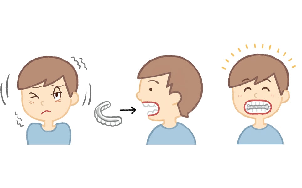
1. An oral splint that can reduce Tourette syndrome tics
A removable dental device that can reduce tics in children and adults with Tourette Syndrome has been developed by scientists at Osaka University.
Biting down on the custom-made oral splint immediately improved both motor and vocal tics in 10 of the 14 children and 6 of the 8 adults who participated in the study. These improvements were still seen after 100 days, especially in patients who were younger when their tics first started.
The researchers aren’t sure exactly how this device reduces tics, but propose that biting down onto the device could act as a sensory trick that alleviates the involuntary tics. The oral splint has the potential to improve the quality of lives of patients with Tourette syndrome.
Alleviating tics
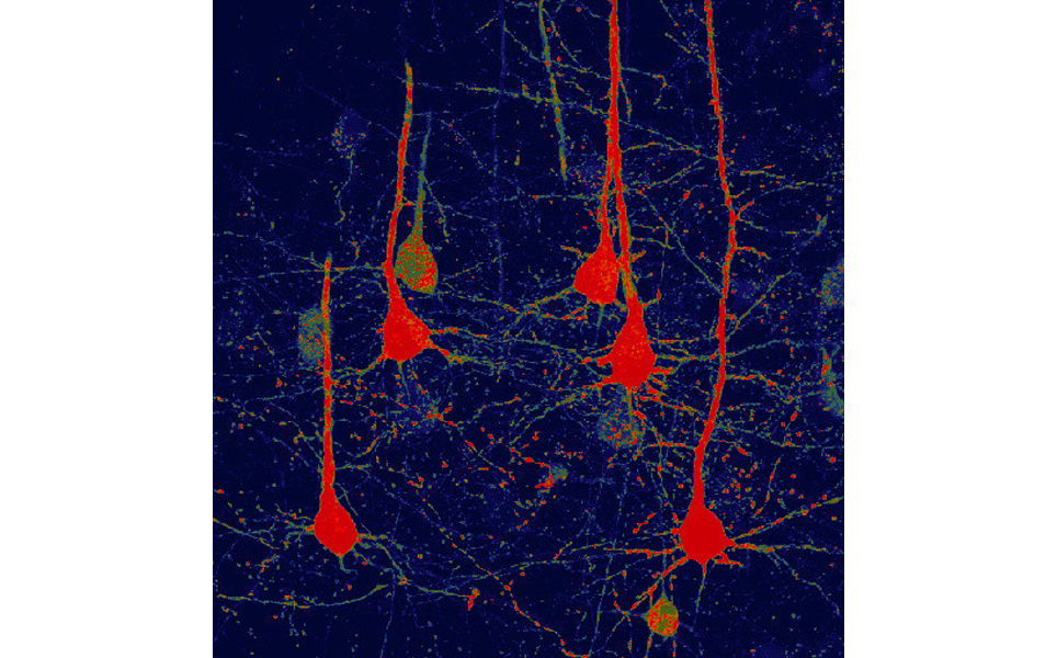
2. Gene therapy may help functional recovery after stroke
A new gene therapy developed by scientists at Penn State University that turns glial cells to functional neurons could improve functional recovery after a stroke.
Glial cells are abundant support cells surrounding every neuron in the brain. Unlike neurons, glial cells are able to regenerate themselves, including after brain injury. This makes them an ideal cell to transform into neurons to replace those that are lost during a stroke.
The gene therapy is based on the NeuroD1 gene, a genetic neural factor that has been previously used to convert glial cells into functional neurons in mouse brains with Alzheimer’s disease. The team used the AAV viral system to deliver NeuroD1 into the motor cortex of a mouse that had suffered a stroke.
Following a stroke, glial cells proliferate and form a glial scar in the affected areas. The AAV system was used to express NeuroD1 in areas that the glial scars had formed, turning these glial cells into neuronal cells. This direct conversion of glial cells to neurons increased the density of neurons in stroke areas and reduced the loss of brain tissue following a stroke.
The newly converted neurons had similar properties to those that were lost due to the stroke; they were fully functional and able to fire action potentials and form synapses. The researchers are next going to further test this method and use it for clinical therapies.
Converting cells to neurons
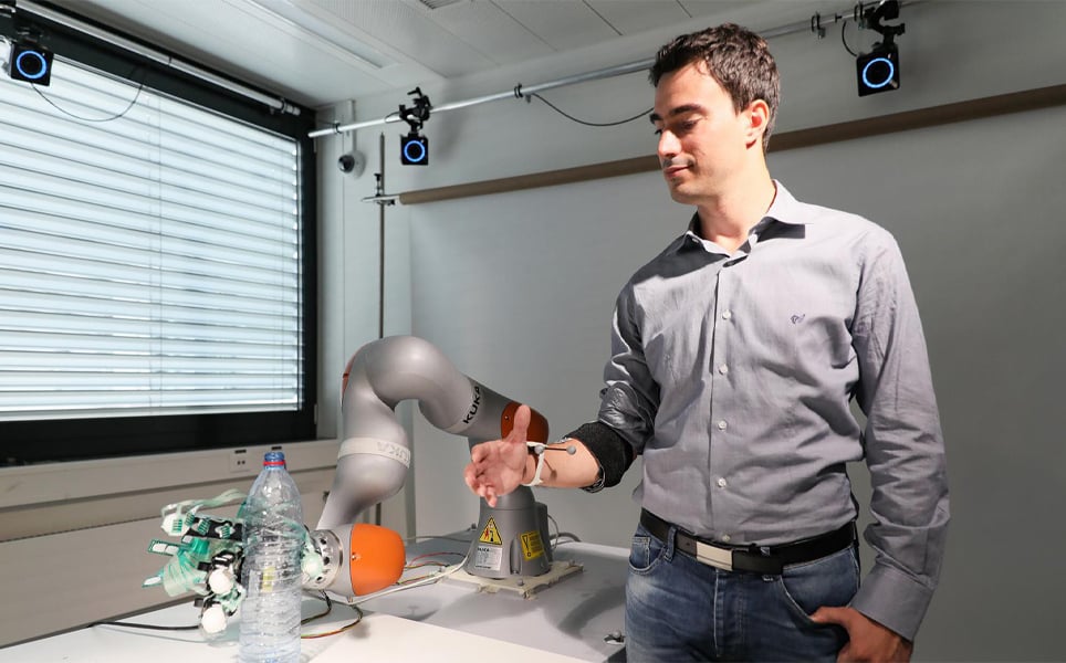
3. A smart artificial hand for amputees merges user and robotic control
A new neuroprosthetic technology that combines robotic control with the users’ voluntary control, giving individual finger control and automation for improved grasping and manipulation, has been developed by scientists at EPFL.
This technology merges neuroengineering and robotics to enable intended finger movement to be deciphered from muscular activity on the amputee’s stump for individual finger control of the prosthetic hand, along with the robotic hand being able to help take hold of objects and maintain contact with them for robust grasping.
The technology uses an algorithm which first learns to decode user intention and translates this into finger movement of the prosthetic hand. To train the algorithm, which uses machine learning, the amputee performs a series of hand movements. Sensors on the amputee’s stump detect muscular activity, and the algorithm learns which hand movements correspond to which patterns of muscular activity, enabling individual fingers on the prosthetic hand to be controlled.
When an object is in contact with sensors on the surface of the prosthetic hand, the algorithm tells the prosthetic hand to close its fingers, enabling automatic grasping.
Find out more
4. Brain tumours form synapses with healthy neurons, Stanford-led study finds
A study by Stanford University has shown for the first time that brain tumours, called high-grade gliomas, form synapses with healthy neurons. This enables them to receive electrical signals that drive their growth.
The high-grade gliomas not only form synapses with healthy neurons, but also contain gap junctions. This means that electrical signals can be transmitted from healthy neurons to the cancerous tissue, and then amplified within the tumours.
The team found that disrupting these signals with an anti-epilepsy drug reduced the ability of the cancer to grow. Perampanel, a seizure medication which blocks activity of synaptic neurotransmitter receptors, reduced proliferation of paediatric gliomas implanted into mice by 50%. Meclofenamate, a drug that blocks the action of gap junctions, also led to a similar decrease in tumour proliferation.
The researchers are going to continue to investigate whether blocking electrical signalling within the tumours could help treat high-grade gliomas.
Cancer cell signalling
5. Baby brain scans made available online to advance research
Brain scans of over 500 newborn babies have been released for researchers to download and use to study the human brain.
The images have been released as part of the Developing Human Connectome Project (dHCP), a collaboration between King’s College, Imperial College London and the University of Oxford, that aims to uncover how the wiring and function of the brain develops during pregnancy and birth. This release of images is the first large-scale release of data of the project.
The images include babies born and imaged between 24 – 45 weeks of pregnancy. Most babies were imaged while naturally asleep. The researchers hope that the data will allow scientists across the world to understand how diseases such as autism develop and how problems in pregnancy affect the brain.
See the brain scans
6. Brain circuit connects feeding and mood in response to stress
A team of researchers led by Baylor College of Medicine have identified a brain circuit in mouse models that connects the feeding and mood centres of the brain.
The team conducted this research to understand the link between depression and other psychiatric disorders and changes in metabolism, including obesity and lack of appetite. They found that POMC neurons in the hypothalamus, essential for regulating feeding behaviour and body weight, have physical connections with another region of the brain that contains numerous dopamine neurons, implicated in the regulation of mood.
Connection between feelings and feeding
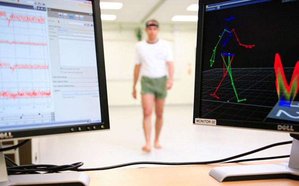
7. For the first time, walking patterns identify specific dementia type
Scientists at Newcastle University have, for the first time, shown that people with Alzheimer’s disease or Lewy body dementia have unique walking patterns that enable the conditions to be distinguished.
The researchers found that people with Lewy body dementia vary their step time and length and are asymmetric when they walk, unlike people with Alzheimer’s disease whose walking patterns are more consistent.
These results suggest that walking could be a useful diagnostic tool for dementia.
New diagnostic tool for dementia
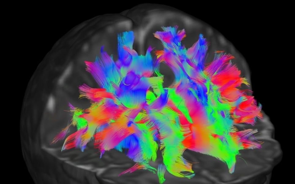
8. Do children’s brains really get thinner?
Scientists at Stanford University and the Max Planck Institute have used brain imaging techniques to find that the brains of children do not thin as they grow as much as expected.
Previous studies have shown that certain regions of the cerebral cortex get thinner as children develop, resulting in as much as 1mm of grey matter being lost by adulthood. Using quantitative MRI, this research suggests that rather than the brains of children thinning as they grow, there is an increase in myelin surrounding nerve fibres.
Myelin is the white matter of the brain and, as measurements of grey matter depend on detecting the border between white and grey matter, the increase in myelin may obscure the measurements of cortical grey matter.
Grey or white matter?
9. AI helps scientists predict depression outcomes
Two studies led by scientists at UT Southwestern suggest that artificial intelligence can be used to identify patterns of brain activity that make a person less responsive to certain antidepressants, enabling treatment outcomes to be predicted.
The studies of more than 300 people used brain imaging to look at brain activity in both a resting state and during the processing of emotions. In both studies, the participants were divided into a healthy control group and patients with depression. Participants either received medication or a placebo.
In the participants who received medication, there was a correlation between how the brain is wired and the likelihood of responding to an antidepressant within two months. Artificial intelligence was used to identify correlations between the effectiveness of an antidepressant and how a patient’s brain processes emotional conflict. AI was able to identify specific brain regions that were most important in predicting whether a patient would benefit from an SSRI antidepressant.
Predicting treatment outcomes
10. Epilepsy: Seizures do not announce themselves as thought
Previously, it was thought that certain patterns of brainwaves preceded an epileptic seizure. These could then be used as indicators to predict when a seizure may occur.
However, scientists at the University of Bonn have found that these changes in patterns of brainwaves, termed ‘critical slowing down’ events, are now unlikely to precede epileptic seizures.
In the study, patients with epilepsy had up to 70 probes measuring the cerebral flow in their brains. The researchers analysed data taken during the hours just before a seizure as well as during a two week period.
The researchers observed a number of critical slowing down events, but found that these usually occurred independently of a seizure. Therefore, using these events as seizure predictors is not reliable and could lead to seizures not only being missed but also being falsely predicted.
Can seizures be predicted?
Banner image credit: The Developing Human Connectome Project
Take a look at the previous top neuroscience stories...
Sign up to receive our latest news
Find out about Scientifica's latest product releases, company news, and developments through a range of news articles, customer interviews and product demonstration videos.
)