Banner image: Confluente Rhabdomyosarcoma (RD) cell line under an inverted phase contrast microscope. Credit: Dhifaf zeki, Wikimedia Commons

What is Phase Contrast?
Phase contrast microscopy is a light microscopy technique used to enhance the contrast of transparent and colourless specimens that would otherwise be difficult to see using conventional brightfield microscopy. Many biological samples, including living cells, absorb very little light and therefore appear almost invisible under a standard microscope. Phase contrast overcomes this by converting small phase shifts in transmitted light into visible differences in image brightness and contrast.
Because phase contrast microscopy does not require cells to be fixed, stained or killed, it allows living cells to be observed in their natural state. This makes it one of the most widely used imaging techniques for cell culture, enabling researchers to monitor cell morphology, growth, migration and division in real time while preserving normal cellular behaviour. Fluorescence microscopy can also be used alongside phase contrast to provide additional structural or molecular information.
Phase contrast is particularly well suited to thin, transparent specimens and is commonly used on inverted microscope systems for imaging cells in culture vessels. The inverted configuration provides additional working space and easier access to samples, although phase contrast can also be implemented on upright microscopes.
For thicker specimens or applications requiring higher resolution and improved edge definition, differential interference contrast (DIC) microscopy may be a more suitable imaging technique.
What is Phase Contrast used for?
Phase contrast microscopy is primarily used to visualise transparent, unstained specimens that produce little natural contrast under conventional brightfield illumination. The technique is particularly valuable for observing living biological samples without the need for fixation or staining, allowing cellular processes to be monitored in real time.
Common applications of phase contrast microscopy include:
- Cell Culture Imaging
- Live Cell Imaging
- Microbiology and Microorganisms
- Thin Tissue Cultures
- Fibres
- Subcellular particles and organelles
How does Phase Contrast work?
Many biological specimens, including living cells, are almost transparent and absorb very little light. As a result, they often appear with poor contrast when viewed using conventional brightfield microscopy. Although these specimens do not significantly alter the intensity of light passing through them, they do change its phase as it travels through regions of different thicknesses and refractive indices.
These unstained specimens are often referred to as phase objects because they primarily affect the phase, rather than the amplitude, of transmitted light. Typically, light diffracted by the specimen is phase-shifted by approximately one-quarter of a wavelength relative to the surrounding background light. While these phase differences contain valuable information about the specimen, they cannot be detected directly by the human eye, which is sensitive only to differences in light intensity and colour.
Phase contrast microscopy converts these otherwise invisible phase differences into visible differences in image brightness and contrast. This allows transparent structures within cells and tissues to be visualised without the need for staining.
To achieve this, light from the microscope illumination source passes through a condenser annulus located within the substage condenser. The annulus produces a hollow cone of illumination that reaches the specimen. Some of this light passes through the specimen without being diffracted, while light interacting with cellular structures is diffracted and undergoes a phase shift.
The undeviated background light is focused into a bright ring at the rear focal plane of the objective, where it aligns with the phase plate. The diffracted light is distributed across the remainder of the objective aperture, allowing the background and specimen-diffracted light to be treated separately.
The phase plate selectively advances or retards the phase of the undeviated background light by approximately one-quarter of a wavelength. Many phase plates also contain a partially absorbing grey ring that reduces the intensity of the background light by around 60–90%, further enhancing image contrast.
When the undeviated and diffracted light waves recombine at the image plane, constructive and destructive interference occur depending on their relative phase. This interaction converts the original phase differences within the specimen into differences in brightness, producing a high-contrast image in which cellular structures become clearly visible against the background.

Figure 1: The path of light in an upright microscope set up for phase contrast.
How to align a microscope for Phase Contrast
For phase contrast microscopy to produce optimal image contrast, the condenser annulus and objective phase ring must be precisely aligned. Misalignment can reduce image quality and contrast, making it more difficult to visualise specimen detail. Although many modern phase sliders and condensers are supplied pre-centred, it is good practice to verify the alignment before use.
1. If possible, set up Köhler illumination on the microscope. See Scientifica’s guide to Köhler illumination for a step-by-step walkthrough.
2. Install the appropriate phase annuli or phase rings in the condenser.
3. Remove one eyepiece and insert the phase contrast centring telescope.
4. Focus the centring telescope until both the condenser annulus and objective phase ring are clearly visible.
5. Select the lowest magnification phase objective and matching condenser annulus. For example, a 10× Ph1 objective should be used with a Ph1 annulus.
6. Observe the condenser annulus and objective phase ring through the centring telescope.
7. Using the condenser centring screws, adjust the condenser annulus until the illuminated ring is concentric with the objective phase ring.

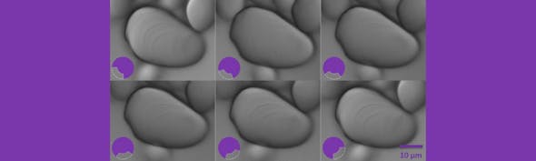
Figure 2: On the left, the objective phase ring and condenser annulus are mis-aligned. On the right, the components are correctly aligned, as the segmented circle of light created by the condenser annulus sits on the black ring.
8. Repeat the alignment procedure for each phase objective and corresponding annulus.
9. Once alignment is complete, remove the centring telescope and replace the eyepiece
Once correctly aligned, phase contrast systems typically require minimal adjustment. However, it is recommended to periodically check alignment using the centring telescope, particularly after servicing, transporting or reconfiguring the microscope.
Advantages of Phase Contrast Microscopy
Phase contrast microscopy offers several advantages for imaging transparent biological specimens, particularly living cells and microorganisms.
Enhanced Contrast Without Staining
Phase contrast converts small differences in the phase of transmitted light into visible differences in image brightness, allowing transparent and colourless specimens to be visualised without the need for dyes or stains.
Imaging of Living Cells
Because samples do not need to be fixed or stained, living cells can be observed in their natural state. This enables researchers to study cellular behaviour while minimising sample preparation and perturbation.
Real-Time Observation of Biological Processes
Phase contrast is well suited to live cell imaging, allowing dynamic processes such as cell growth, migration, division and morphological changes to be monitored over time.
Simple and Rapid Sample Preparation
Unlike many fluorescence-based techniques, phase contrast typically requires little or no sample preparation, making it ideal for routine laboratory observation and quality control of cell cultures.
Cost-Effective Imaging
Phase contrast does not require fluorescent labels, specialised probes or additional reagents, making it a cost-effective technique for many routine imaging applications.
Compatibility with Other Imaging Techniques
Phase contrast can be combined with fluorescence microscopy, enabling researchers to visualise overall cell morphology while simultaneously imaging specific molecular targets.
Widely Applicable
Phase contrast is commonly used for imaging cultured cells, microorganisms, protozoa, embryos and other thin, transparent specimens across a wide range of biological and biomedical applications.
Limitations of Phase Contrast Microscopy
While phase contrast is a powerful technique for imaging transparent specimens, it also has several limitations that should be considered when selecting an imaging method.
Halo Artefacts
One of the most common limitations of phase contrast microscopy is the appearance of halo artefacts around specimen edges. These bright outlines can obscure fine structural details and make image interpretation more difficult.
Reduced Performance with Thick Specimens
Phase contrast performs best with thin, transparent samples. Thicker specimens can scatter light more strongly, reducing image quality and introducing unwanted artefacts.
Limited Resolution Compared to DIC
Although phase contrast provides excellent contrast, it generally offers lower image resolution and less precise edge definition than differential interference contrast (DIC) microscopy.
Quantitative Measurements Can Be Challenging
The brightness variations produced by phase contrast are related to optical path length rather than directly representing physical structures, which can make quantitative image analysis more difficult.
Specialised Optical Components Required
Phase contrast microscopy requires dedicated condenser annuli and phase objectives that must be correctly matched and aligned to achieve optimal image quality.
Not Suitable for All Samples
Highly scattering, strongly absorbing or very thick specimens may be better imaged using alternative contrast techniques such as DIC, fluorescence microscopy or confocal microscopy.
Phase Contrast vs DIC
A comparison table of Phase Contrast vs Differential Interference Contrast (DIC)

Learn more about other contrast techniques

Scientifica PatchScope
An ultra-stable, motorised inverted microscope that can be integrated with your existing manipulators.