

In Vivo Patch Clamp Electrophysiology: Methods, Applications and Practical Insights

by Scientifica
Introduction
As neuroscience recording technologies continue to evolve, researchers have more options than ever for studying brain activity. High-density silicon probes can record from thousands of neurons simultaneously, advanced imaging approaches can visualise activity across entire populations, and emerging voltage indicators promise new ways of measuring neuronal dynamics.
Yet despite these advances, one technique continues to hold a unique place in the neuroscientist’s toolkit:
in vivo whole-cell patch clamp electrophysiology.
In a recent webinar hosted by the British Neuroscience Association in partnership with Scientifica, Dr Rebecca Jordan and Dr Soraya Meftah from the University of Edinburgh explored why in vivo patch clamp recordings remain one of the most powerful approaches available for investigating neuronal computation in the intact brain.
Their message was clear: if you want to understand not just whether a neuron fires, but why it fires, few techniques can match the insight provided by intracellular recordings.
Why study neuronal function?
At its core, neuroscience seeks to understand how billions of neurons work together to produce thoughts, behaviours, memories and perceptions.
Every one of these processes depends on electrical communication between neurons. Some of this activity occurs at synapses, where neurons exchange information. Some appears as action potentials—the spikes that serve as the primary output signal of neurons. At larger scales, populations of neurons generate oscillations and network activity that coordinate brain function across regions.

Slide courtesy of Dr Soraya Meftah, University of Edinburgh
During the webinar, Dr Meftah used a memorable football stadium analogy to illustrate these different levels of organisation. Imagine a packed stadium containing thousands of spectators. Individual conversations between fans are analogous to local neuronal communication. Larger coordinated chants, waves or crowd responses resemble network-level activity occurring across brain regions.
Different recording techniques allow researchers to listen to different parts of this activity. Standing outside the stadium might tell you that something exciting is happening, but not who scored the goal. Sitting beside a single fan allows you to hear individual conversations but provides only a narrow perspective.
In many ways, electrophysiology faces a similar challenge. Researchers must decide whether they want to study populations, networks or individual neurons - and select the appropriate technique accordingly.
What makes whole-cell recordings different?
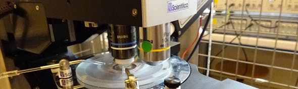
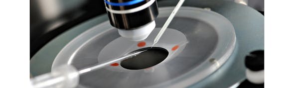
Whole-cell patch clamp electrophysiology provides direct access to the interior of a neuron.
Using a fine glass micropipette filled with an intracellular solution, researchers establish a high-resistance seal with the neuronal membrane before gaining electrical access to the cell.
Once whole-cell configuration is achieved, the experimenter can directly measure:
- Membrane potential
- Synaptic inputs
- Action potential generation
- Intrinsic excitability
- Ion channel function
- Receptor-mediated signalling
- Cellular responses to experimental manipulations
Unlike extracellular recording techniques, which detect electrical signals outside neurons, whole-cell recordings reveal the subthreshold activity occurring inside the cell.
This distinction is critical.
Many of the computations performed by neurons occur before an action potential is generated. Excitatory and inhibitory inputs are integrated, filtered and transformed into output activity through processes that are often invisible to extracellular recording methods.
Whole-cell recordings allow researchers to observe these computations directly.
Choose the right micromanipulator for your in vivo electrophysiology setup
From multielectrode recordings to in vivo patch clamp, selecting the right micromanipulator is critical for stable recordings, precise targeting, and efficient workflows.
This helpful guide will help you identify the right setup for your application.

Why perform patch clamp recordings in vivo?
Traditional whole-cell recordings are commonly performed in cultured neurons or acute brain slices. These preparations provide excellent experimental control and remain essential tools for neuroscience research.
However, they also remove neurons from many of the network interactions that shape their behaviour in the living brain.
In vivo patch clamp recordings preserve those interactions.
By recording from neurons in the intact brain, researchers can investigate how individual cells behave while receiving natural sensory inputs, participating in ongoing network activity and contributing to behaviour.
As Dr Meftah explained, this makes in vivo patch clamp particularly valuable when researchers want to understand how neurons function within complete circuits rather than in isolation.

Slide courtesy of Dr Soraya Meftah, University of Edinburgh
The approach is especially powerful for studying:
- Sensory processing
- Sensorimotor integration
- Brain-state dependent activity
- Network oscillations
- Learning and memory
- Neurological disease
- Behavioural computation
Blind patching vs two-photon targeted patching
Modern in vivo patch clamp experiments typically use one of two major approaches.
Blind patch clamp recordings
Blind patching relies on monitoring changes in electrode resistance as the recording pipette advances through brain tissue.
Rather than directly visualising neurons, the experimenter identifies cells by detecting characteristic resistance changes that occur when the pipette approaches a membrane.
The process requires considerable technical skill.
Researchers must carefully control pipette pressure, monitor resistance measurements and interpret subtle electrical signals to determine when they have encountered a neuron.
As Dr Jordan described during the webinar, success often depends on developing an intuitive understanding of what is happening at the pipette tip despite being unable to see it directly.
The technique remains widely used because it offers several practical advantages:
- Simpler optical requirements
- Compatibility with deep brain structures
- Reduced imaging complexity
- Flexibility across experimental preparations
Two-photon targeted patch clamp
For researchers who require visual targeting, two-photon microscopy provides a powerful alternative.
A commonly used method known as shadow patching involves including a fluorescent dye within the pipette solution. As the pipette advances through tissue, dye enters the extracellular space, causing neurons to appear as dark shadows against a fluorescent background.
This enables researchers to target specific neurons directly while simultaneously visualising surrounding structures.
The advantages include:
- Cell-specific targeting
- Morphological reconstruction
- Simultaneous imaging
- Easier dual recordings
- Integration with calcium imaging
The result is a uniquely rich dataset that combines physiology and imaging in a single experiment.
What can in vivo patch clamp reveal?
The real power of in vivo patch clamp lies in the questions it can answer.
During the webinar, both speakers presented examples where whole-cell recordings revealed phenomena that would have been difficult to detect using other methods.

Slide courtesy of Dr Soraya Meftah, University of Edinburgh
Understanding sensory processing in dementia
Dr Meftah discussed work investigating sensory processing in a mouse model of dementia.
Previous studies characterising this model had demonstrated abnormalities in neuronal responsiveness using calcium imaging in vivo, in addition to identified reductions in synaptic proteins and excitatory receptor expression. However, when conventional in vitro electrophysiology was performed, many expected functional differences were not apparent.
This raised an important question.
Were there key differences to how a neuron integrates information in vivo which weren’t captured in vitro? To answer this, researchers turned to in vivo whole-cell recordings in the somatosensory cortex.
By recording responses to whisker stimulation in the intact brain, they were able to examine how individual neurons integrated sensory information under natural network conditions.
The recordings revealed differences in sensory integration that were not fully captured by slice electrophysiology alone, demonstrating how circuit context can fundamentally alter neuronal function.
The study highlights one of the key strengths of in vivo patch clamp: the ability to bridge the gap between cellular physiology and network behaviour.
Detecting prediction errors in visual cortex
Dr Jordan presented a second example that illustrates another unique advantage of whole-cell recordings.
A central challenge faced by the brain is distinguishing sensory inputs generated by our own actions from those originating in the external world.
When we move our eyes, turn our heads or walk through an environment, we create large amounts of self-generated sensory input. Yet we are still able to identify unexpected events occurring around us.
One proposed solution is that the brain computes prediction errors.
In this framework, neurons compare expected sensory consequences of movement with actual incoming sensory information. Any mismatch generates a prediction-error signal that highlights unexpected events.
To test this idea, Dr Jordan’s group performed whole-cell recordings from neurons in the visual cortex of awake mice.

Slide courtesy of Dr Rebecca Jordan, University of Edinburgh
The researchers simultaneously measured locomotion-related signals and visual motion signals while recording membrane potential.
They discovered neurons that integrated these two sources of information with opposing signs. In effect, the neurons appeared to subtract expected visual consequences of movement from actual visual input.
Importantly, these computations were visible primarily in subthreshold membrane potential signals.
Had the researchers relied solely on spike recordings, much of this information would likely have remained hidden.
This example demonstrates why whole-cell recordings continue to occupy a unique niche within modern systems neuroscience.
Key to success in in vivo patch clamping
In vivo patch clamp is often regarded as one of the most technically demanding techniques in neuroscience. Success depends on many variables, and recording quality can be influenced by factors that might seem insignificant at first glance.
The speakers highlighted several recurring themes.
High quality micropipettes
The micropipette serves as the primary interface between the experimenter and the neuron.
Its geometry, resistance and cleanliness all influence recording success.
For somatic recordings, pipettes in the range of approximately 5–7 MΩ are commonly used, with smooth tip geometry helping to maximise seal formation.
Reliable pressure control
Pressure regulation is essential throughout the recording process.
Different stages require different pressure levels, from maintaining pipette cleanliness during brain entry to searching for neurons and forming stable seals.
Without this, recordings would have low success.
Mechanical stability
Perhaps the greatest enemy of successful recordings is movement.
Even micron-scale shifts between the brain and pipette can disrupt a recording.
Stable head fixation systems and robust mechanical design therefore play a central role in successful experiments.
Surgical Technique
Good recordings often begin long before the pipette enters the brain.
Clean craniotomies, refined surgical procedures and careful tissue handling all contribute to improved success rates.
Training and troubleshooting
Both speakers emphasised the importance of experience.
Learning to interpret resistance changes, identify technical issues and troubleshoot complex setups takes time.
In many laboratories, developing expertise in patch clamp electrophysiology is measured in months or years rather than weeks.
In vivo patch clamp vs Neuropixels
One audience question focused on how whole-cell recordings compare with modern silicon probe technologies such as Neuropixels.
The answer, according to both speakers, depends entirely on the scientific question.
Neuropixels probes provide extraordinary recording density and can capture activity from hundreds or thousands of neurons simultaneously.
For studies focused on:
- Population activity
- Circuit interactions
- Spike timing
- Large-scale recordings
they are often the ideal choice.
However, Neuropixels recordings remain extracellular.
They cannot directly reveal membrane potential dynamics, synaptic integration or many forms of subthreshold computation.
Whole-cell recordings remain unmatched when researchers need to understand how individual neurons transform inputs into outputs.
Rather than competing technologies, the two approaches should be viewed as complementary tools that answer different questions.
Limitations to consider
Despite its strengths, in vivo patch clamp is not the right solution for every experiment.
Researchers must balance its advantages against several important limitations.
These include:
- Low throughput compared with extracellular methods
- Limited recording durations
- Sensitivity to movement
- Technical complexity
- Challenges associated with voltage clamp recordings in vivo
- Increased difficulty when targeting deep brain structures
For many questions, simpler approaches may provide sufficient information.
The key is selecting the method that best aligns with the scientific objective.
Looking ahead
As new technologies emerge, the landscape of neuronal recording continues to evolve.
Genetically encoded voltage indicators and advanced optical methods are beginning to provide access to membrane potential signals at scales previously impossible.
Yet for now, in vivo whole-cell recordings remain the benchmark for studying single-neuron physiology in the intact brain.
For researchers seeking to understand how neurons integrate information, participate in circuits and generate behaviour, patch clamp electrophysiology continues to provide a level of mechanistic insight that few other methods can match.
And while the technique demands patience, technical expertise and perseverance, the scientific rewards remain substantial.
Dr Rebecca Jordan
Principal Investigator, University of Edinburgh
Dr Rebecca Jordan is Principal Investigator and Simons ESAT Fellow at the University of Edinburgh, where she leads the Prediction and Plasticity Lab within the Centre for Discovery Brain Sciences. Her research focuses on how the brain learns to predict sensory input and integrate sensory and motor information to support perception and behaviour. She uses a combination of cutting-edge systems neuroscience techniques, including in vivo electrophysiology, two-photon imaging, optogenetics, and virtual reality paradigms, to investigate cortical circuit function and learning mechanisms.

Dr Soraya Meftah
Dr Soraya Meftah is a Postdoctoral Research Fellow at the UK Dementia Research Institute and Centre for Discovery Brain Sciences at the University of Edinburgh. Her research focuses on the neurophysiology of dementia, investigating synaptic, neuronal, and network dysfunction in neurodegenerative diseases such as Alzheimer’s. She has extensive experience in both in vivo and in vitro patch clamp electrophysiology, alongside advanced techniques including imaging and molecular approaches. Her work combines high-resolution electrophysiology with translational disease models, including studies using live human brain tissue, to better understand early circuit dysfunction in neurodegeneration.

This blog was developed using AI based on content from the webinar and has been reviewed for accuracy. If you notice any issues, please contact [email protected].


)