

Using Scientifica electrophysiology equipment for multipatch experiments to investigate Dravet syndrome
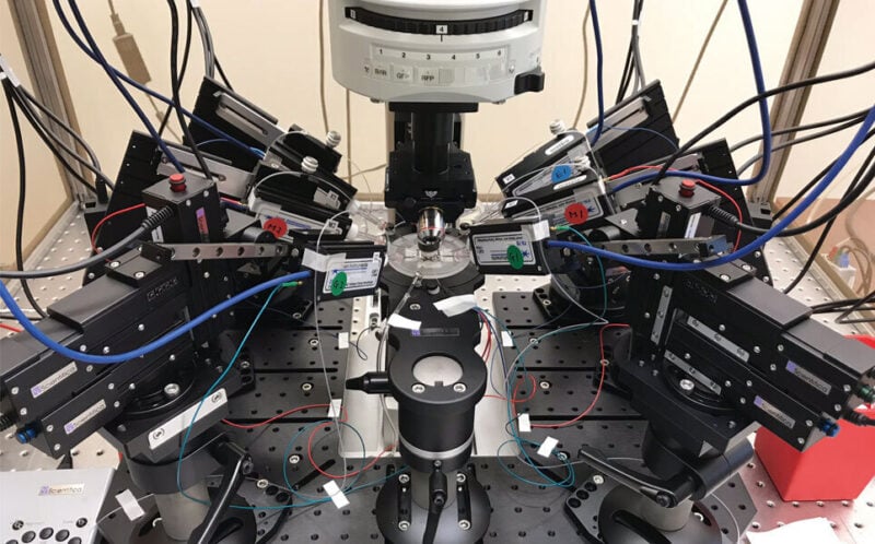
A recent journal publication in Cell Press from the laboratory of Ethan Goldberg at The Children's Hospital of Philadelphia, uses the Scientifica SliceScope Pro 6000 with four Scientifica PatchStar Micromanipulators for multi-electrode electrophysiology recordings.
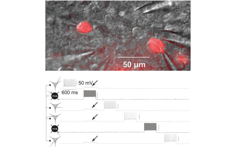
The goal of the Goldberg NeuroLab is to develop new treatments and work towards a cure for epilepsy and other neurodevelopmental disorders, using a range of innovative approaches in experimental systems. This particular study uses a mouse model of Dravet syndrome and interrogates how the mutation modifies synaptic transmission and action potential propagation in fast spiking interneurons. The investigators utilise a novel approach called "mini-slices" to perform multi-patch experiments on the same animal at two timepoints to measure how the mutation delays signal transfer.
About the Study
Dravet syndrome is a neurodevelopmental disorder characterized by epilepsy, intellectual disability, and sudden death due to pathogenic variants in SCN1A with loss of function of the sodium channel subunit Nav1.1. Nav1.1-expressing parvalbumin GABAergic interneurons (PV-INs) from young Scn1a+/− mice show impaired action potential generation. An approach assessing PV-IN function in the same mice at two time points shows impaired spike generation in all Scn1a+/− mice at postnatal days (P) 16–21, whether deceased prior or surviving to P35, with normalization by P35 in surviving mice. However, PV-IN synaptic transmission is dysfunctional in young Scn1a+/− mice that did not survive and in Scn1a+/− mice ≥ P35. Modeling confirms that PV-IN axonal propagation is more sensitive to decreased sodium conductance than spike generation. These results demonstrate dynamic dysfunction in Dravet syndrome: combined abnormalities of PV-IN spike generation and propagation drives early disease severity, while ongoing dysfunction of synaptic transmission contributes to chronic pathology.
Learn More
A word from the Goldberg NeuroLab
"The SliceScope Pro 6000 has a lot of flexibility; the system is compact with a wide workspace thus compatible for multiple cell patching. This means there is an ultra-high degree of freedom to make setting up the manipulators easier. Moreover, the motorised base plate and multiple manipulators are very stable and smooth. These are the reasons why I am happy I chose the SliceScope Pro 6000 to perform multiple cell patching."
Dr Keisuke Kaneko, Children's Hospital of Philadelphia
Scientifica SliceScope Pro 6000
The SliceScope Pro 6000 is an integrated electrophysiology system with a static microscope. The incredible versatility of the system means that you can effortlessly switch between in vitro and in vivo electrophysiology experiments, and perform advanced imaging, such as two-photon imaging and confocal microscopy.
Scientifica PatchStar Micromanipulator
The perfect electrophysiology micromanipulator – from single channels to in vivo field recordings. The stable design and ultra-quiet electronics mean you can perform long-term patch clamp experiments and record even the smallest signals.
Paper Reference
Kaneko, K., Currin, C. B., Goff, K. M., Wengert, E. R., Somarowthu, A., Vogels, T. P. & Goldberg, E. M. (2022). Developmentally regulated impairment of parvalbumin interneuron synaptic transmission in an experimental model of Dravet syndrome. Cell Press, 38(13). https://doi.org/10.1016/j.celrep.2022.110580
Banner credit: Goldberg NeuroLab