
Neuron-microglia communication in the CNS after epileptic seizures
The means by which neurons regulate microglia are not well understood. However, recent research has illuminated the mechanism by which neurons control morphological changes in microglia after induced seizure activity.
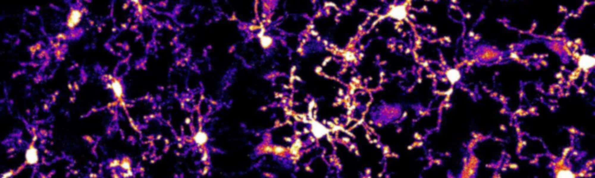
Seizures are associated with a significant increase in extracellular glutamate, causing a spike in neuronal activity. These periods of intense neural activity also lead to alterations in the shape of microglia, as visualised in a study published in The Journal of Neuroscience. The images were captured by two-photon time-lapse microscopy on Scientifica’s Multiphoton Imaging System.
These results led researchers to investigate the role of glutamate in these modifications. To do this they monitored microglia in the CA1 region of the hippocampus using high resolution two-photon microscopy of brain slices. Application of glutamate led to an increase in the amount of microglial process extension. Additionally, the same treatment had a similar effect in cortical slices and in vivo.
With further investigation the group led by Dr Long-Jun Wu from Rutgers University managed to elucidate the molecular mechanism that leads to the increase in microglial process extension. They found that glutamate released during increased neural activity opens calcium channels on post-synaptic membranes through the NMDA receptor. This intracellular calcium influx causes a release of ATP from the post-synaptic neuron which is detected by P2Y12 receptors on the microglial processes, promoting growth towards increasing concentrations of ATP. The mechanism of release of ATP is currently unknown although ion channels like pannexin 1 or pre-packaged vesicles are likely candidates.
The scientists also looked at P2Y12 KO mice. The P2Y12 receptor is necessary for process extension during neuronal hyperactivity, as shown by a lack of morphological change in these mice. Furthermore, P2Y12 KO mice showed worse levels of seizures when induced through delivery of Kainic Acid (KA). This suggests that microglia have a neuro-protective function during periods of hyperactivity.
Hopefully, future studies will be able to show how this neuro-protective function operates and may lead to a therapeutic strategy against disorders such as epilepsy.
)