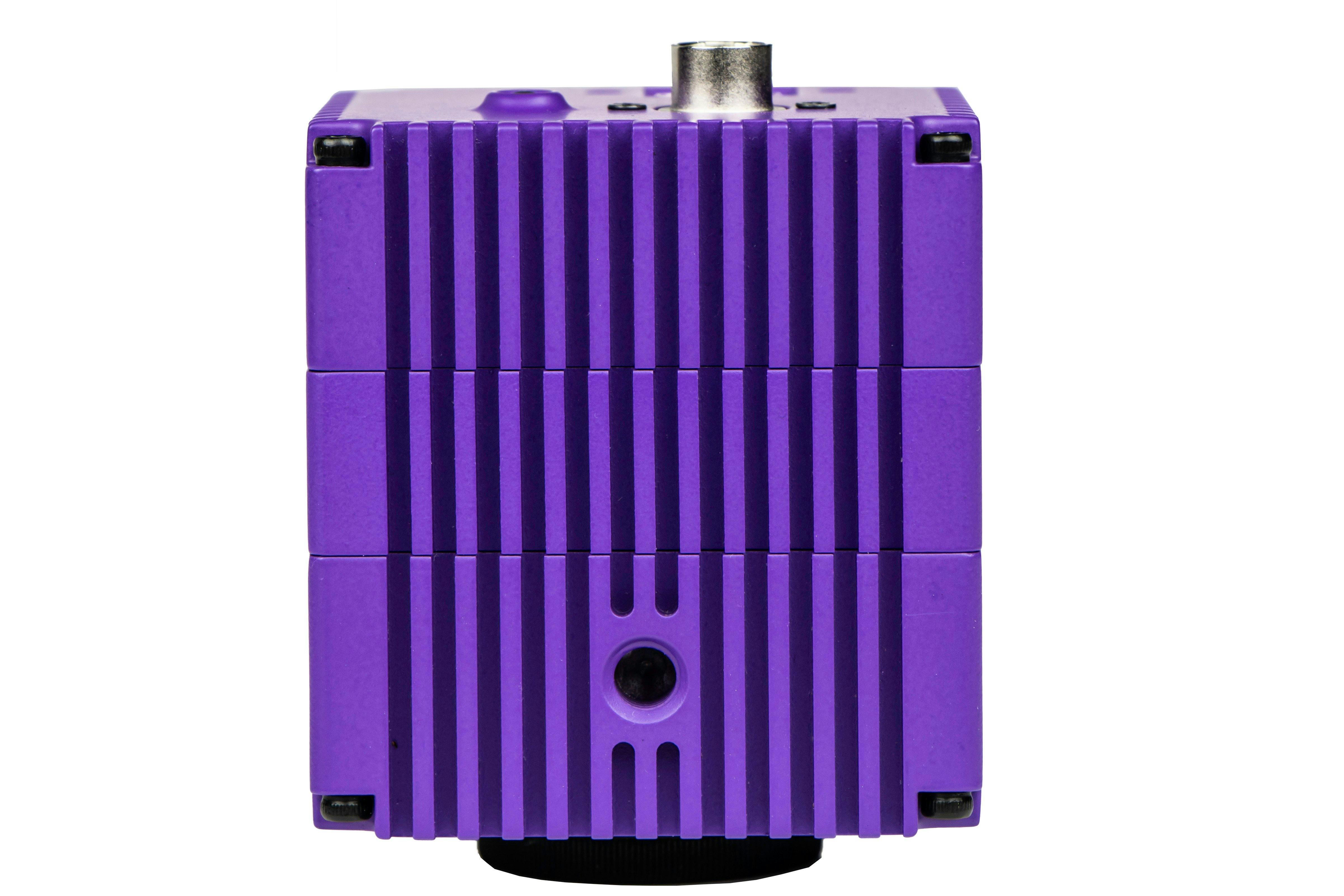
Scientifica SciCam+ Camera
Visualise high-contrast cells for precise patch clamping and fluorescent imaging, with seamless data capture
The SciCam+ is a high-resolution sCMOS camera purpose-built to meet the demands of patch clamp electrophysiology. Its fast frame rate and high sensitivity allow for precise electrode positioning and clear visualisation of even the smallest cellular structures.
Ideal for Infrared (IR) Differential Interference Contrast (DIC) imaging, as well as fluorescence, live cell, and calcium imaging, the SciCam+ delivers high contrast and customisable settings, ensuring accurate data collection and smoother experimental workflows.
Product benefits
Testimonials
Images acquired using Scientifica the SciCam+ camera
Mouse hippocampus was visualised using IR-DIC on the SciCam+ at 40x objective magnification. Sample courtesy of Vincenzo Mastrolia, Kings College London


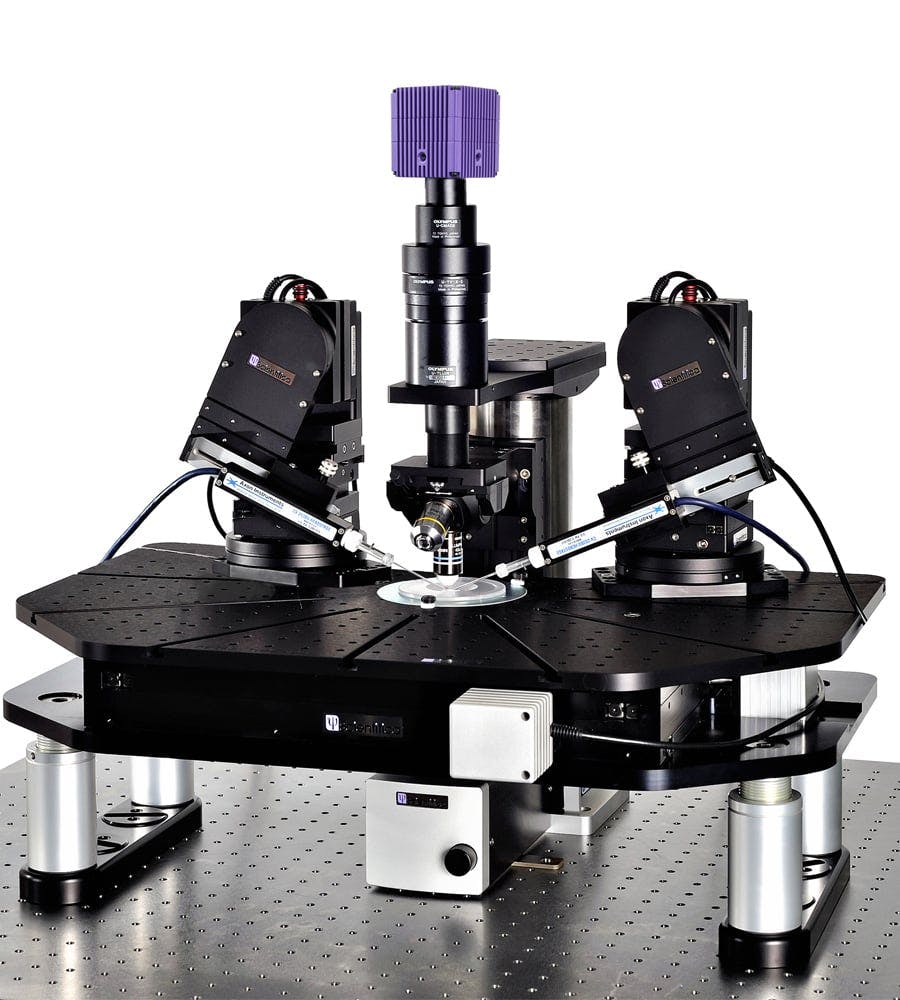
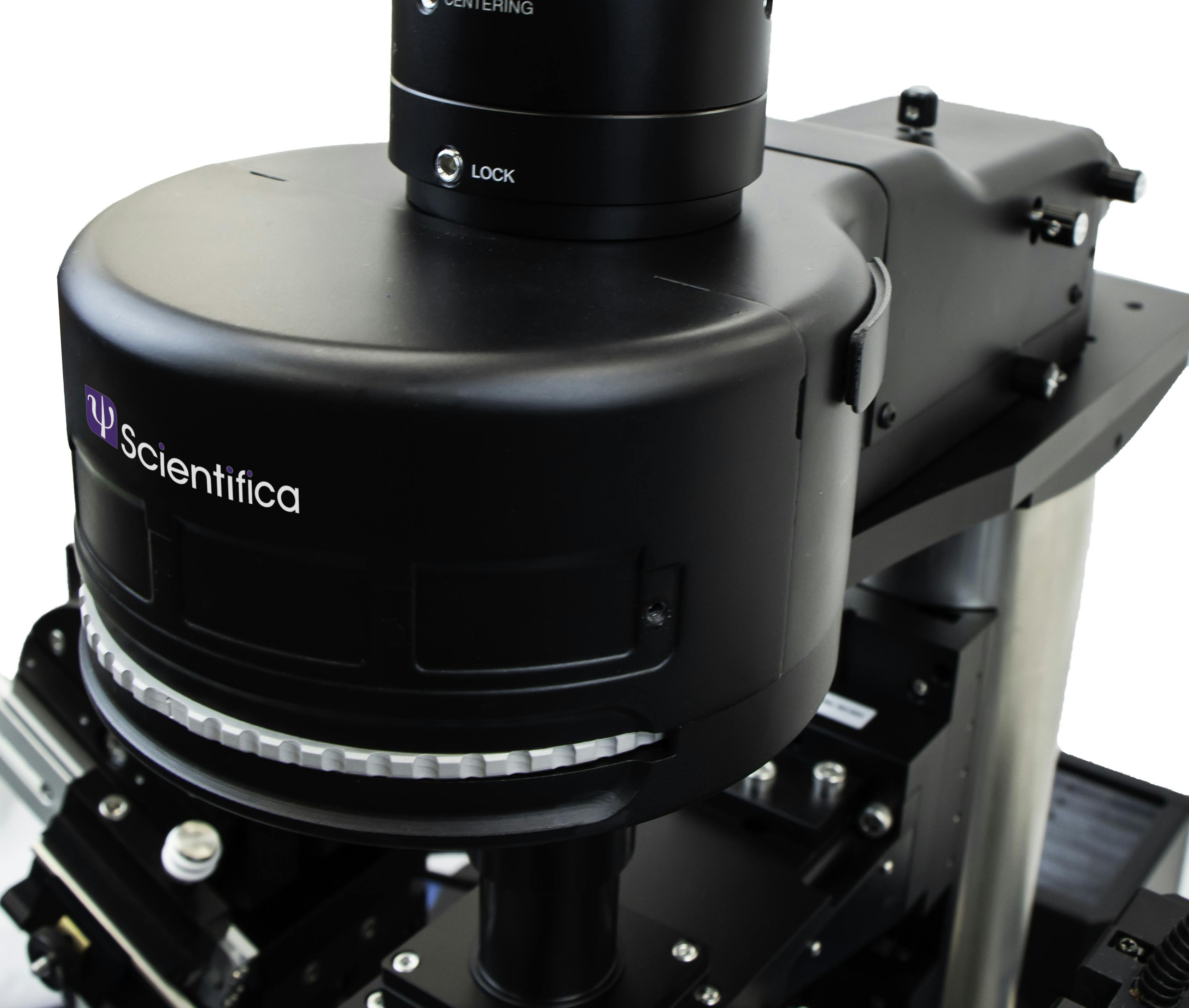
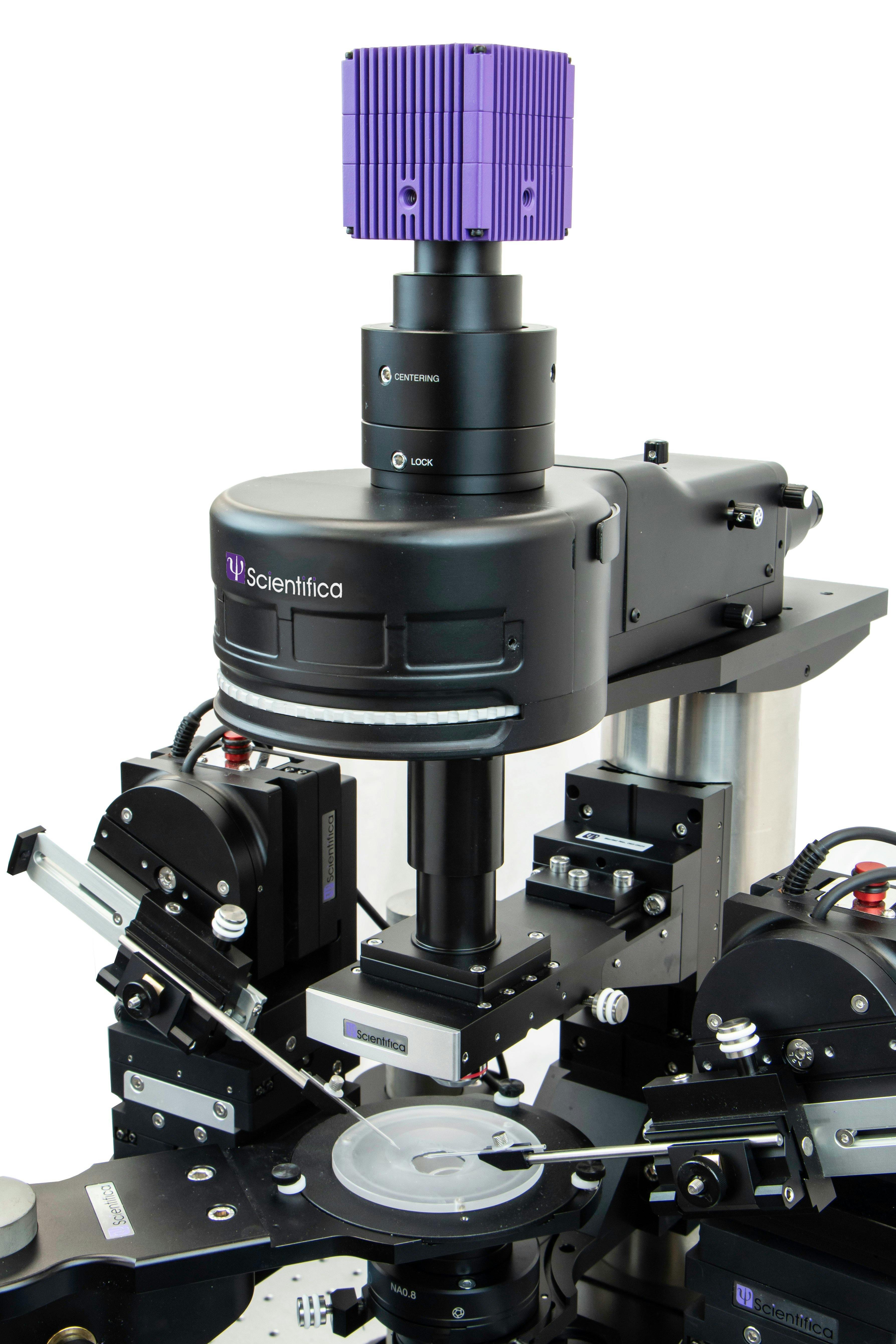
Scientifica SciCam+ on patch clamp rig - SliceScope Pro 1000
Applications
- Patch Clamp
- Electrophysiology
- Tissue slice electrophysiology
- IR-DIC imaging
- Fluorescence imaging
- Calcium imaging
- Live cell imaging
Scientifica services for this product
Speak to one of our experts for details on pricing, features, installation and support.