

Scientifica's picks of the top neuroscience stories from February 2020
Our selection of the best neuroscience stories published in February include the discovery of how a childhood brain tumour grows and proliferates, a new non-invasive way to measure axons and harnessing the brain's immune cells to improve memory.
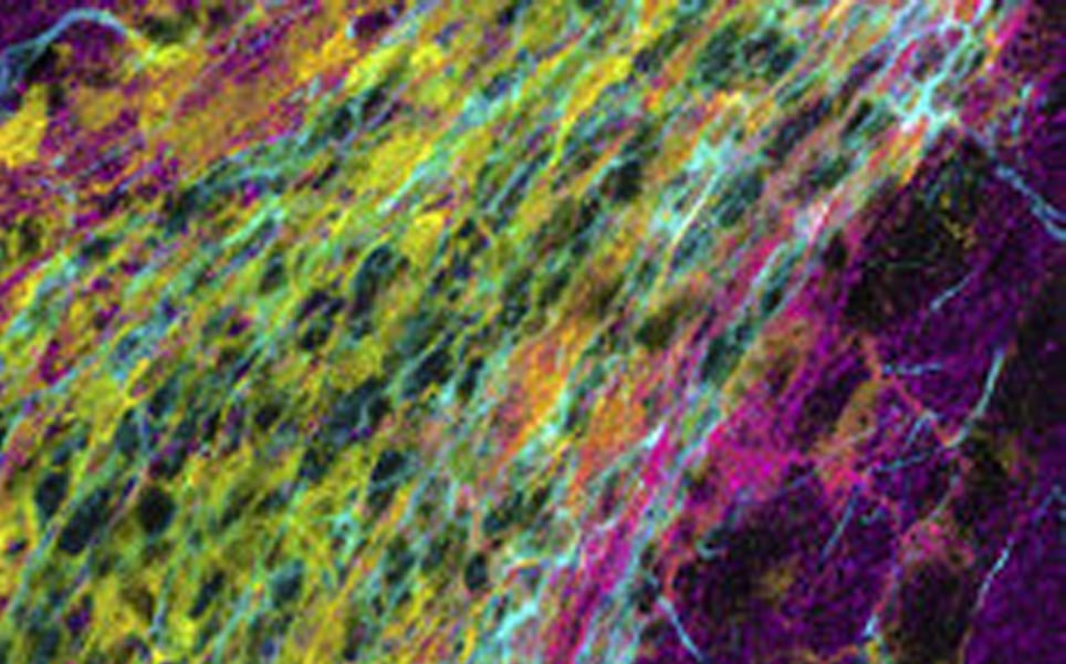
1. Brain tissue stiffness is crucial for neurogenesis
Neural stem cells are only present in a few specific parts of the adult mammalian brain. Only these parts of the brain, called niches, are capable of generating new neurons. Researchers at Helmholtz Zentrum München have characterised the proteome of these niches for the first time, and compared this to the proteome of other regions of the brain to identify the factors that enable these niches to generate new neurons.
The scientists found that the proteome of the niche encodes an extracellular matrix with a high solubility. It also encodes transglutaminase 2, a multi-functional enzyme that ensures strong cross-links. The team found that transglutaminase 2 is essential for neurogenesis regulation, and it is thought that it may contribute to the stiffness of the stem cell niche in otherwise soft brain tissue.
Next, the researchers want to compare these proteomes to the environment following brain injury, with the aim of converting the injured region into a neurogenic stem cell niche, to create new neurons after injury.
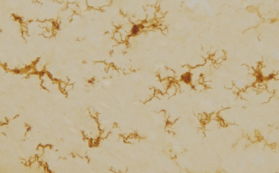
Neurogenic niche
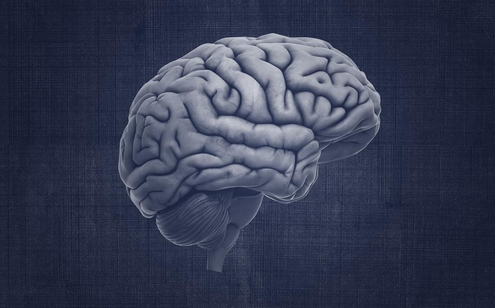
2. Childhood brain tumour discovery may unlock new treatments for many cancers
It has been discovered that a childhood brain cancer, which has been studied by researchers at the University of Virginia for its supposed simplicity, forms an unexpectedly intricate network to drive its growth.
In a model of the cancer, the researchers marked medulloblastoma tumour cells green. They found that all cells surrounding the tumour were colourless, but astrocytes were green. As these astrocytes shared their green colour with tumour cells, it suggested that they originated from the tumour cells, meaning that some tumour cells change their identity to form a new cell type. The team found that these astrocytes hijack microglia and cause it to produce insulin-like growth factor 1, a substance that is essential for tumour proliferation.
The researchers now hope that they can target this intricate network to stop tumour proliferation.
Understanding tumour proliferation
3. A smart jumpsuit provides information on infants’ movement and development
A smart jumpsuit that accurately measures the spontaneous and voluntary movement of infants from the age of five months has been developed by researchers at the University of Helsinki.
This enables the movements of infants outside the lab to be quantified, helping neurological development abnormalities to be detected and assessed earlier. It is thought that in the future, this smart jumpsuit can be used to measure how various therapies and treatments affect children’s development.

Innovative jumpsuit
4. Antidepressant harms baby neurons in lab-grown ‘mini-brains’
Lab-grown ‘mini-brains’, which are derived from stem cells and act as models of the developing brain, have been used by scientists at Johns Hopkins University to investigate the effects of the common antidepressant paroxetine on the developing brain.
Paroxetine is able to cross the placenta and currently comes with a warning against its use in early pregnancy. The researchers found that it suppresses the growth of synapses between neurons and causes significant decreases in oligodendrocyte support-cells. This suggests that paroxetine may hinder the normal formation of interconnections between developing neurons, which could underlie autism or other disorders.
As the mini-brains are made from human cells, they may be able to better predict the effects of substances on the brain. They can also be mass-produced in the lab, making them a cheaper alternative to animal models.
More about the mini-brains
5. Brain asymmetry is not unique to humans
It has long been thought that asymmetry between the left and right hemispheres of the brain is unique to humans. However, scientists at the Max Planck Institute for Evolutionary Anthropology and the University of Vienna have found that chimpanzees, gorillas and orangutans have the same asymmetry pattern.
Using endocasts, which are imprints of the brain on cranial bones, the researchers were able to see that the magnitude of asymmetry was similar in humans and most great apes, with chimpanzees having less asymmetry than humans, gorillas and orangutans.
Asymmetry of human brains had the largest variation around the most common pattern, which the researchers believe is a sign of the increased functional and developmental modularisation of the human brain that occurred through evolution.
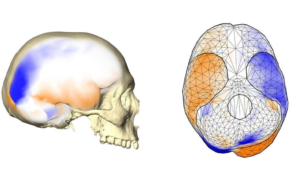
Shared brain asymmetry between species
6. Scientists are locating new brain coordinates of facial blindness
Researchers at the University of Copenhagen have used brain scans to try to map regions of the brain that go awry in those with facial blindness.
People with facial blindness are unable to recognise the features that make our faces unique and so can’t recognise different faces.
The team used brain scans to compare the brain activity of 15 adults who are face-blind with 33 control subjects. It was expected that an area known as the ‘fusiform face area’ on the right side of the brain would show different activity in face-blind individuals, as this area processes visual impressions when looking at faces. However, activity in this region was similar in both groups, but differed in a similar area of the left hemisphere. The researchers found that brain activity in this region of the left hemisphere was impaired in face-blind individuals compared to control subjects.
As so little is known about the causes of facial blindness, understanding which areas of the brain are affected is a step closer to developing therapies to help those who suffer from it.

Advancing understanding of facial blindness
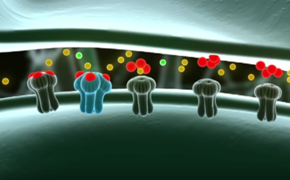
7. MRI method provides unprecedented insight into the brain’s wiring network
In a collaborative study, researchers at YU Grossman School of Medicine, Champalimaud Centre for the Unknown and the Cardiff University Brain Research Imaging Centre (CUBRIC) have found a way to non-invasively measure axons.
The diameter of axons is only a few micrometres, making it impossible, up until now, to non-invasively measure them. The thickness of axons affects their conduction of neural impulses.
The researchers developed a method that uses magnetic resonance imaging (MRI), and separates signals originating inside the axons from those arising from the surrounding tissue. Using signals originating in the axons, the researchers were able to measure their properties. Measurements were more accurate than ever before, with only a 10 - 15% error.
Next, the researchers would like to measure axon diameters over extended periods, to help understand how they change over time in both health and diseases.
Find out more
8. Antibodies: the body’s own antidepressants
Researchers at the Max Planck Institute of Experimental Medicine in Göttingen have found that autoantibodies may act as the body’s own antidepressants.
After investigating the mechanisms that cause autoantibodies that target the NMDA glutamate receptor to be generated, the researchers discovered that the level of these autoantibodies in the blood can increase during chronic stress. They found that people suffer less depression and anxiety when these antibodies are able to act on NMDA receptors in the brain, suggesting they are acting as antidepressants.
Autoantibody antidepressants
9. How the brain’s immune system could be harnessed to improve memory
Immune cells in the brain are often associated with neurodegenerative diseases that affect memory. However, researchers at RMIT have found that microglia can be activated to have the reverse effect and improve memory, rather than cause it to deteriorate.
The researchers observed up to a 50% improvement in performance of memory tasks when microglia were altered in rats. Although the effect was temporary, the researchers hope that the discovery will help with the development of new therapies aimed at enhancing memory formation and preventing age-related cognitive decline.
Harnessing microglia to improve memory
10. McGill researchers end decade-long search for mechanical pain sensor
Scientists at McGill University have discovered that the TACAN protein, which is found in sensory neuron membranes, is involved in detecting mechanical pain.
The function of the TACAN protein has previously been unclear. The researchers have shown for the first time that the protein forms ion channels in the membrane of pain-sensing cells, where it converts mechanical pressures into electrical signals, enabling mechanical pain to be detected.
In a mouse model, when TACAN was switched off, the animals were significantly less sensitive to mechanical pain stimuli.
This finding could enable the development of new, powerful analgesic drugs that target this protein, which could be used to treat conditions such as osteoarthritis and neuropathic pain.
Learn more
Banner image credit: Simon Neubauer, CC BY-NC-ND 4.0
Take a look at the previous top neuroscience stories...
Sign up to receive our latest news
Find out about Scientifica's latest product releases, company news, and developments through a range of news articles, customer interviews and product demonstration videos.
)