

Using a SliceScope to combine calcium imaging and laser scanning photostimulation
Tuan D. Nguyen, Physics Department, The College of New Jersey
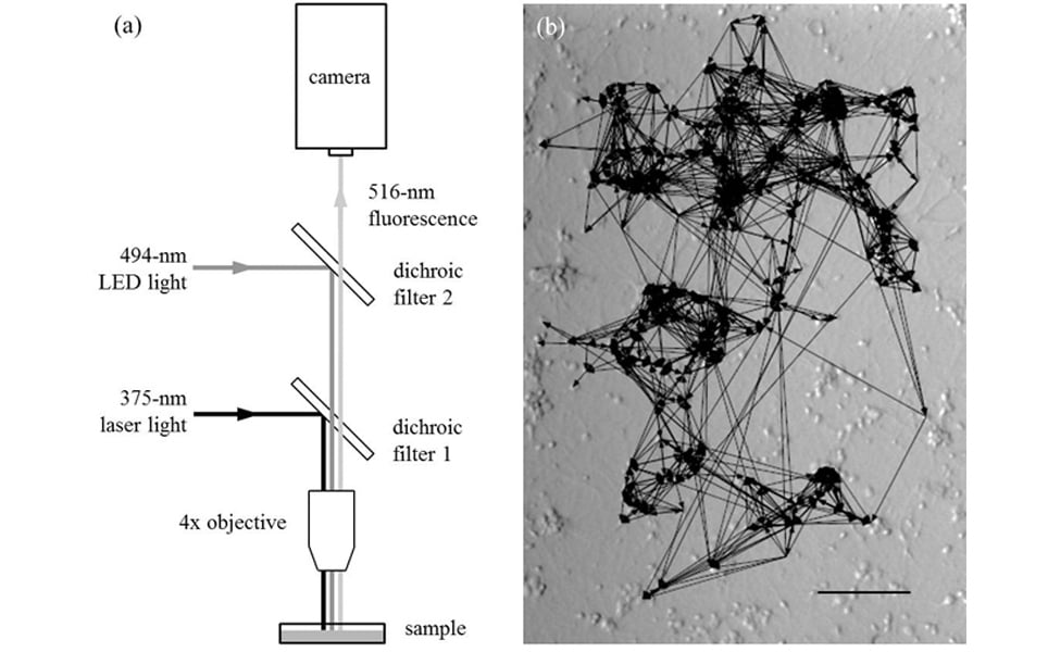
My research focuses on understanding the neural activity of a large population (100-200) of neurons based on the underlying connectivity. To do this, I needed a system that can simultaneously stimulate neurons while monitoring their activity. Laser scanning photostimulation (LSPS) [1] and calcium (Ca) imaging [2] were two well-known, all-optical techniques at the time (2012). Unfortunately, at the time, they could only be done separately. To perform Ca imaging, it was possible to add additional components to the existing microscope system. For LSPS, components do not exist so it was necessary to make modifications to the microscope itself, as I did when I was a postdoc.
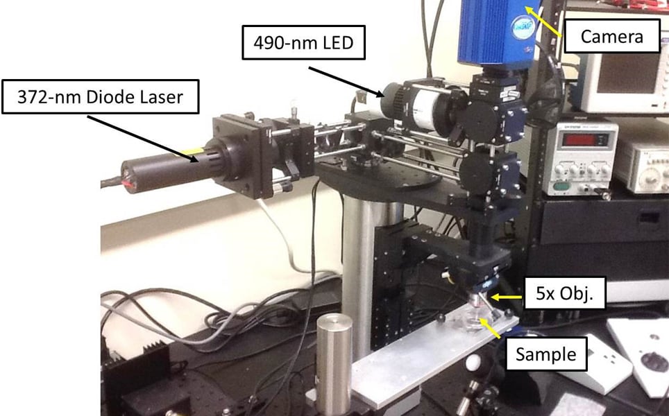
The SliceScope was the ideal system for what I wanted to do. It was modular and straightforward enough that it was easy to add components. The schematic below shows how it was done. Using galvo-driven mirrors, the system allows stimulation of any neuron within the field-of-view (FOV) whilst looking for Ca responses from postsynaptic neurons. In this way, a connectivity map of the neuronal network consisting of about 200 neurons can be made within 15 minutes. We have used this system to study network synchronisation in cultured neurons [3] and will continue these investigations.
In addition, we would like to make the system even more versatile by adding contrast illumination and patch-clamp electrophysiology components. This would allow us to study neural microcircuits in brain slices using both optical and electrophysiological techniques.
Scientifica SliceScope
A stable and compact upright microscope. The modular design makes the SliceScope a perfect foundation for many different configurations including in vivo or in vitro electrophysiology, multiphoton imaging and confocal imaging.
References:
- L. C. Katz and M. B. Dalva, “Scanning laser photostimulation: a new approach for analyzing brain circuits.,” J. Neurosci. Methods, vol. 54, no. 2, pp. 205–218, Oct. 1994.
- Y. Ikegaya, M. Le Bon-Jego, and R. Yuste, “Large-scale imaging of cortical network activity with calcium indicators.,” Neurosci. Res., vol. 52, no. 2, pp. 132–138, Jun. 2005.
- T. D. Nguyen, K. D. O’Connor, K. Sheth, and N. Bolle, “Mapping functional connectivity of bursting neuronal networks,” Appl. Netw. Sci., vol. 2, no. 15, pp. 1–10, Jun. 2017.
Find out about Scientifica's latest product releases, company news, and developments through a range of news articles, customer interviews and product demonstration videos: