

Using a Scientifica Multiphoton Microscope for investigating the neural circuitry underlying behaviour
Dr. Michael Bale, Scientifica and University of Sussex
Research has come a long way in understanding how science can use animal models to understand the brain and behaviour. Mice are now trained to perform fairly complex behavioural tasks that are designed to interrogate how neural circuits process events in their environment, relate to their previous experience and then act accordingly. Many tasks have been adapted to have the mouse stationary while still allowing it free movement virtually on a ball, treadmill or in a home-cage environment. This approach has been taken advantage of by the scientific community so that activity in the brain can be measured via electrophysiology or imaged with advanced microscopy techniques while an animal receives a stimulus, makes a decision and takes an appropriate action.
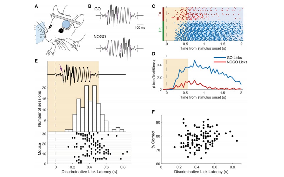
In one such study from the University of Sussex, researchers investigated how neurons of the primary somatosensory cortex could distinguish between different sequences of vibrations. Mice were trained to receive sensory stimuli delivered to their whiskers while being head-fixed underneath a Scientifica MP-2000 multiphoton microscope. These mice also expressed a calcium indicator, GCaMP6f, so the activity of several neurons could be visualised at once while the animal performed the task. Mice were able to receive fluid rewards upon reporting that they had felt a target vibration by licking.
The primary somatosensory cortex is described in all textbooks for principally representing sensory information from the outside world. In the rodent whisker system this is primarily indicated by curvature (bending) of whiskers exerting a force in follicles in the skin. However, contrary to the doctrine, in animals trained in a sequence discrimination task, the researchers found almost no neurons in the somatosensory cortex that could discriminate between stimulus categories, but discovered that many neurons actually responded to the mice making decisions or licking. In naïve mice, not previously exposed to the task, neurons did show some sensory selectivity. But neurons did not show any action-related responses even when they licked for a reward on correct trials. In subsequent behavioural sessions the number of action-related neurons increased with experience.
A future goal of systems and behavioural neuroscience will be to tease apart the roles of individual cell subtypes that allow the brain to process information over short (seconds) and long (days) timescales. One way this could be achieved is to perform longitudinal imaging of a brain region of interest and employ optogenetic tools.
Two-photon in vivo imaging experiments were conducted using Scientifica’s Multiphoton Galvo System, equipped with a pulsed infrared laser. Wavelengths of 940 nm were used to excite GCaMP6f in cortical layers II–III. Image acquisition, triggered externally, was controlled by ScanImage (Vidrio) at a resolution of 256 × 100 pixels at 10.8 frames per second using galvo scanners. Custom-made behavioural apparatus were mounted on a Scientifica post and platform fixed to a motorised moveable base plate (MMBP).
Paper reference
Bale, M., Bitzidou, M., Giusto, E., Kinghorn, P., Maravall, M. Sequence Learning Induces Selectivity to Multiple Task Parameters in Mouse Somatosensory Cortex. Current Biology (2020) doi: https://doi.org/10.1016/j.cub.2020.10.059