

Looking without seeing: Exogenous attentional guidance from primates to fish
By Veronique van den Berghe
Professor Li Zhaoping of the Department of Computer Science at University College London, recently gave a seminar at the Centre for Developmental Neurobiology at King’s College London as part of the ‘NEUReka!’ seminar series 2017-2018. She explained how the brain creates saliency maps for what the retina sees and how the map for exogenous attentional guidance has ‘migrated’ from the optic tectum to the primary visual cortex during evolution.
Vision is composed of three stages: encoding, (attentional) selection and decoding. During the encoding stage, retinal input light is converted into neuronal activity. As our brain has limited resources to process what our eyes continuously transmit, only a fraction of the visual input is selected. This results in an unconscious blindness to non-selected inputs. Finally, during the decoding stage we recognise the object from the selected inputs. In other words, selection and decoding correspond to looking and seeing, respectively.
This raises the questions: How does our brain decide which visual inputs to select and process before we have seen anything? Where does this crucial selection take place?
Attention helps to focus the limited processing power to a fraction of the visual input during the selection stage. The guidance of our gaze is either endogenous or exogenous. Internal factors, such as the goal of an ongoing task, voluntarily guide our attention in an endogenous or top-down manner. Knowledge of the environment and input features, such as colour or shape, increase the efficiency of top-down visual selection. On the other hand, selection by goal-independent mechanisms driven by external visual inputs is essential to promptly respond to unexpected events such as sudden danger. Exogenous guidance by these external factors is involuntary and hence bottom-up. Saliency of a visual location, as defined by Prof. Zhaoping, is the degree to which a location attracts exogenous attention and is measured by the brevity of the reaction time necessary to gaze or saccade towards this location (see also Fig. 1).
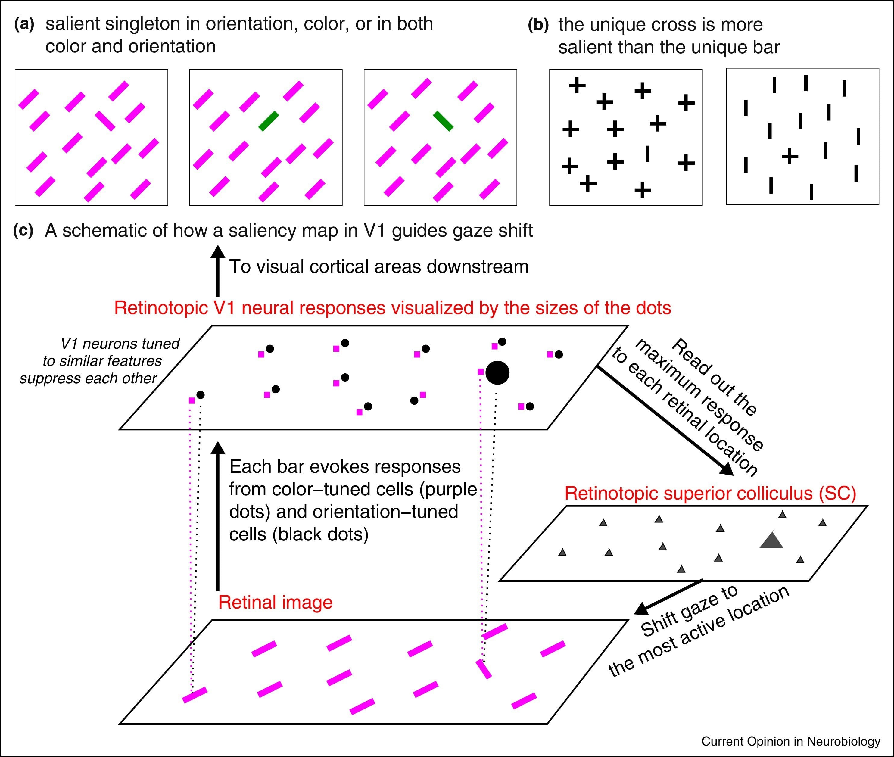
a, b Illustrative examples of visual search. The search target in (a) is a left tilted and/or coloured bar whereas the ease of the search changes by swapping identities of targets and non-targets (b).
c Bottom-up saliency of a location is represented by the maximum response of the primary visual cortex (V1) to this location. The simplified V1 only contains two kinds of neurons, one tuned to colour (purple dots) and the other tuned to orientation (black dots). Each input bar evokes responses in a cell tuned to its colour and another cell tuned to its orientation. Iso-feature suppression makes nearby V1 neurons tuned to similar features (e.g., similar colour or similar orientation) suppress each other and hence the orientation singleton evokes the highest V1 response resulting in a gaze shift to the salient location.
Reprinted from “From the optic tectum to the primary visual cortex: migration through evolution of the saliency map for exogenous attentional guidance” by L. Zhaoping, 2016, 40:94-102.
In primates, the frontal eye field (FEF) of the prefrontal cortex is associated with endogenous attentional guidance. In lower vertebrates and non-mammals which lack a neocortex, such as fish, the superior colliculus (SC) or the optic tectum (OT) respectively guide visual attention. OT and/or SC neurons in fish, amphibians and birds are tuned to orientation and/or motion direction and further enable to compute saliency. Primate SC cells show very little or no specificity for patterns whereas neurons in the primary visual cortex (V1) do.
Hence, in contrast to the traditional view, Prof. Zhaoping suggests that in primates a bottom-up saliency map is created in V1 rather than in the FEF. This indicates that exogenous and endogenous attentional guidance are steered by different brain areas. The migration of the saliency map along the vertebrate phylogenetic tree is further supported by the increased importance for retinothalamic pathways over retinotectal pathways in primates, as well as the projection areas of the retinal ganglion cells in the brain and the effects of lesions in V1 or SC/OT.
A large part of our cerebral cortex is devoted to visual function and thus understanding visual processing in primates as well as in other animals is crucial. Understanding vision will give us insight into how the brain works, or as Francis Crick appropriately said: “There is no scientific study more vital to man than the study of his own brain. Our entire view of the universe depends on it.”
Scientifica supports the NEUReka! Seminar Series to help them book speakers from top research institutions around the world.
Banner: Culture of rat brain cells stained with antibody to MAP2 (green), Neurofilament (red) and DNA (blue) by Gerry Shaw under Creative Commons Attribution-Share Alike 3.0 Unported license. Resized from the original
)